Lymphoepithelial cyst of the pancreas mimicking malignant cystic tumor: report of a case
- Affiliations
-
- 1Department of Surgery, Chungbuk National University, College of Medicine and Medical Research Institute, Cheongju, Korea. jwchoi@chungbuk.ac.kr
- 2Department of Pathology, Chungbuk National University, College of Medicine and Medical Research Institute, Cheongju, Korea.
- 3Department of Radiology, Chungbuk National University, College of Medicine and Medical Research Institute, Cheongju, Korea.
- KMID: 2043290
- DOI: http://doi.org/10.14701/kjhbps.2015.19.3.129
Abstract
- Lymphoepithelial cysts of the pancreas are a type of true cyst that can mimic pseudocysts and cystic neoplasms. They are very rare, non-malignant lesions that are unilocular or multilocular cystic lesions lined predominantly by mature squamous epithelium and surrounded by non-neoplastic lymphoid elements. We, herein, present a patient with a cystic pancreas tumor mimicking a malignant cystic neoplasm. The patient was admitted with upper abdominal discomfort. Computed tomography showed a 64x39 mm cystic mass in the pancreas tail. She underwent distal pancreatectomy and splenectomy. In the fluid analysis of the pancreas cystic mass, the CEA and CA19-9 were 618 ng/ml and 3.9 U/ml, respectively. The resected pancreas specimen showed a 6.5 cm-sized cyst the pancreas tail. The cyst was well circumscribed and multilocular. The final pathology report of the resected pancreas specimen noted that the cyst was multilocular, and the cyst lining was showing stratified squamous epithelium covering the lymphoid tissue (containing lymphoid follicles), which was consistent with a lymphoepithelial cyst. The patient recovered uneventfully from surgery and has been doing well for the past 3 months. A differential diagnosis of cystic pancreatic lesions is important. We suggest that lymphoepithelial cysts, although very rare, may be included in the differential diagnosis of cystic pancreatic tumors.
Keyword
MeSH Terms
Figure
-
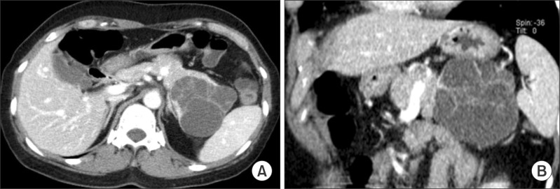
Fig. 1 Preoperative computed tomography images: Axial (A) and coronal (B) contrast-enhanced images show a large cystic mass with internal septa in the tail of the pancreas.
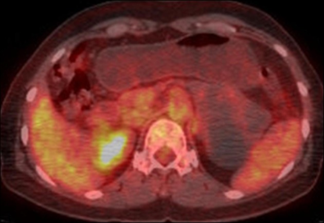
Fig. 2 The transaxial FDG PET-CT image shows no abnormal FDG tracer uptake at the pancreatic cystic mass.
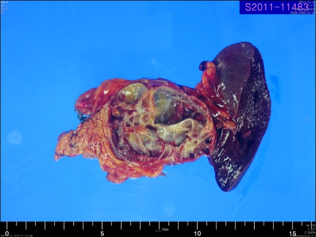
Fig. 3 A photograph of the gross specimen shows a 6.5 cm-sized lymphoepithelial cyst of the pancreas tail. The cyst is well circumscribed and multilocular.
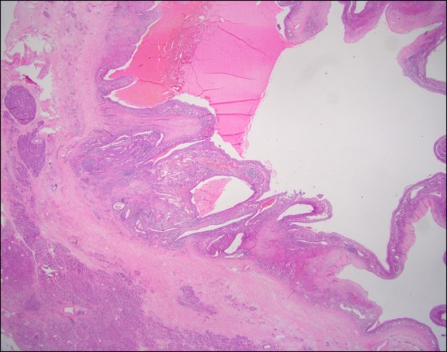
Fig. 4 A low-power view filed of the lymphoepithelial cyst. The cyst is multilocular, and the normal pancreatic parenchyma is seen in the lower left corner (H&E, ×40).
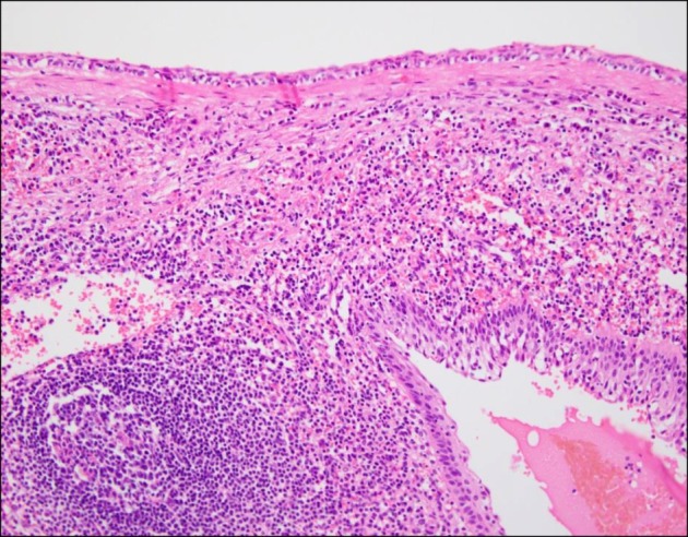
Fig. 5 A high-power view filed of the cyst-lining shows stratified squamous epithelium covering lymphoid tissue containing a lymphoid follicle (H&E, ×200).
Reference
-
1. Brugge WR, Lauwers GY, Sahani D, Fernandez-del Castillo C, Warshaw AL. Cystic neoplasms of the pancreas. N Engl J Med. 2004; 351:1218–1226. PMID: 15371579.
Article2. Lüchtrath H, Schriefers KH. A pancreatic cyst with features of a so-called branchiogenic cyst. Pathologe. 1985; 6:217–219. PMID: 4048076.3. Osiro S, Rodriguez JR, Tiwari KJ, Rodriguez II, Mathenge N, Tubbs RS, et al. Is preoperative diagnosis possible? A clinical and radiological review of lymphoepithelial cysts of the pancreas. JOP. 2013; 14:15–20. PMID: 23306330.4. Basturk O, Coban I, Adsay NV. Pancreatic cysts: pathologic classification, differential diagnosis, and clinical implications. Arch Pathol Lab Med. 2009; 133:423–438. PMID: 19260748.
Article5. Visser BC, Yeh BM, Qayyum A, Way LW, McCulloch CE, Coakley FV. Characterization of cystic pancreatic masses: relative accuracy of CT and MRI. AJR Am J Roentgenol. 2007; 189:648–656. PMID: 17715113.
Article6. Terakawa H, Makino I, Nakagawara H, Miyashita T, Tajima H, Kitagawa H, et al. Clinical and radiological feature of lymphoepithelial cyst of the pancreas. World J Gastroenterol. 2014; 20:17247–17253. PMID: 25493042.
Article7. Kim WH, Lee JY, Park HS, Won HJ, Kim YH, Choi JY, et al. Lymphoepithelial cyst of the pancreas: comparison of CT findings with other pancreatic cystic lesions. Abdom Imaging. 2013; 38:324–330. PMID: 22610041.
Article
