Clear Cell Adenocarcinoma of the Uterine Cervix in a 15-Year-Old Girl: A Case Report
- Affiliations
-
- 1Department of Radiology, Gachon University Gil Hospital, Incheon, Korea. jekim625@hanmail.net
- KMID: 2041955
- DOI: http://doi.org/10.3348/jksr.2013.69.4.321
Abstract
- Cervical cancer is rare in the pediatric population. In cases of cervical cancer, adenocarcinoma is predominantly reported. Clear cell adenocarcinoma (CCAC) of the uterine cervix is a very rare tumor and accounts for only 4% of all adenocarcinomas of the uterine cervix. Risk factors and pathogenesis of this disease are not exactly revealed. The intrauterine exposure to diethylstilbestrol (DES) and associated non-steroidal estrogen during pregnancy before 18 weeks is the only known risk factor. This study reports the imaging finding of primary uterine cervical tumor in a 15-year-old girl, who was finally diagnosed with CCAC, with no maternal history of DES exposure in utero.
MeSH Terms
Figure
-
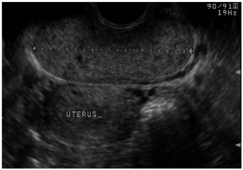
Fig. 1 A 15-year-old adolescent with clear cell adenocarcinoma of the cervix. Axial transrectal pelvic ultrasonograph shows a 7 × 3 cm well defined homogeneous intermediate echoic mass in the upper vagina and cervix.
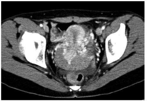
Fig. 2 Axial contrast-enhanced CT image demonstrates a 7 × 4 cm well defined homogeneous, enhancing mass in the upper vagina and cervix.
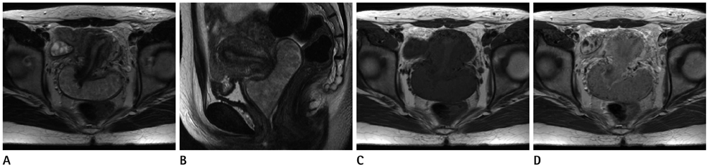
Fig. 3 Axial (A) and sagittal (B) T2-weighted images show a lobulated homogeneous, hyperintense mass in the upper vagina and cervix. No parametrial extension is evident and interfaces between the mass, bladder, and rectum are sharply delineated without evidence of tumor invasion. (C) Non-contrast axial T1-weighted image shows an isointense mass versus myometrium. (D) Gadolinium enhanced axial T1-weighted image shows a homogeneous, well-enhanced mass.
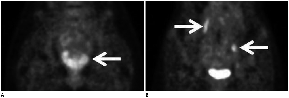
Fig. 4 A. F-18 fluorodeoxyglucose positron emission tomography/CT image shows hypermetabolic changes in the upper vagina and cervix (arrow). B. Hypermetabolic change in right common iliac and left external iliac area is noted (arrows).
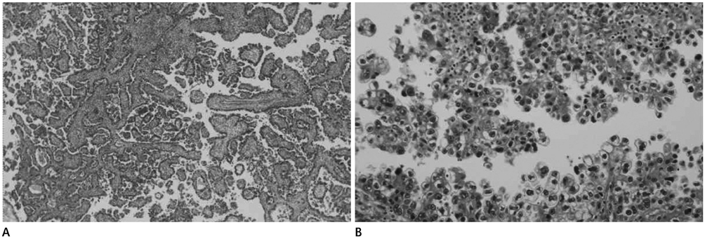
Fig. 5 Pathologic features of clear cell adenocarcinoma of the cervix. Photomicrograph shows infiltrating tumor cells with a tubular-cystic growth pattern in cervical stroma. Tumor cells with clear cytoplasm often project into the lumen of cysts and tubules with a 'hobnail appearance'. A. H&E, × 40. B. H&E, × 200.
Reference
-
1. McNall RY, Nowicki PD, Miller B, Billups CA, Liu T, Daw NC. Adenocarcinoma of the cervix and vagina in pediatric patients. Pediatr Blood Cancer. 2004; 43:289–294.2. Herbst AL, Ulfelder H, Poskanzer DC. Adenocarcinoma of the vagina. Association of maternal stilbestrol therapy with tumor appearance in young women. N Engl J Med. 1971; 284:878–881.3. Hanselaar A, van Loosbroek M, Schuurbiers O, Helmerhorst T, Bulten J, Bernhelm J. Clear cell adenocarcinoma of the vagina and cervix. An update of the central Netherlands registry showing twin age incidence peaks. Cancer. 1997; 79:2229–2236.4. Alipour P, Arjmandi K, Hallaji F. Vaginal clear cell adenocarcinoma with early pulmonary metastasis in a child. Pediatr Hematol Oncol. 2008; 25:679–684.5. Donnelly LF, Gylys-Morin VM, Warner BW, Hillard PJ. Case report: clear cell adenocarcinoma of the vagina in a 5-year-old girl: imaging findings. Clin Radiol. 1998; 53:69–72.6. Herbst AL. Clear cell adenocarcinoma and the current status of DES-exposed females. Cancer. 1981; 48:2 Suppl. 484–488.7. Singh P, Nicklin J, Hassall T. Neoadjuvant chemotherapy followed by radical vaginal trachelectomy and adjuvant chemotherapy for clear cell cancer of the cervix: a feasible approach and review. Int J Gynecol Cancer. 2011; 21:137–140.8. Diaz JP, Sonoda Y, Leitao MM, Zivanovic O, Brown CL, Chi DS, et al. Oncologic outcome of fertility-sparing radical trachelectomy versus radical hysterectomy for stage IB1 cervical carcinoma. Gynecol Oncol. 2008; 111:255–260.9. Agrons GA, Wagner BJ, Lonergan GJ, Dickey GE, Kaufman MS. From the archives of the AFIP. Genitourinary rhabdomyosarcoma in children: radiologic-pathologic correlation. Radiographics. 1997; 17:919–937.10. Liu QY, Huang L, Lin XF, Li HG, Gao M, Liang BL. Clinical manifestations and MRI features of vaginal endodermal sinus tumors in four children. Pediatr Radiol. 2013; 43:983–990.
- Full Text Links
-
- Actions
-
Cited
- CITED
-
- Close
- Share
-
- Similar articles
-
- A case of clear cell adenocarcinoma in the uterine cervix of 52-year-old virgin
- A case of clear cell adenocarcinoma of the uterine cervix in a 6-year-old girl
- Cytology of the Uterine Cervico-vaginal Smear of Clear Cell Adenocarcinoma in Uterine Cervix: Report of a Case
- A case of clear cell carcinoma that is unrelated to diethystilbestrol of the uterine cervix
- A Case of Clear Cell Adenocarcinoma in the Uterine Cervix of 22 Years-Old Virgin
