Sarcomatoid Carcinoma of the Small Intestine: A Case Report and Review of the Literature
- Affiliations
-
- 1Department of Radiology, Sanggye Paik Hospital, Inje University College of Medicine, Seoul, Korea.
- 2Department of Radiology, Haeundae Paik Hospital, Inje University College of Medicine, Busan, Korea. radyjh@hanmail.net
- 3Department of Pathology, Sanggye Paik Hospital, Inje University College of Medicine, Seoul, Korea.
- KMID: 2041950
- DOI: http://doi.org/10.3348/jksr.2013.69.4.295
Abstract
- Sarcomatoid carcinomas are rare biphasic tumors composed of mixed malignant epithelial and mesenchymal cells. A few cases for small intestinal sarcomatoid carcinoma were reported. Moreover, most of the cases are focused on the pathologic review. We experienced a case of monophasic sarcomatoid carcinoma arising in the jejunum in a 78-year-old man. In this case, CT showed fungating mass with central necrosis in the jejunum. We also reviewed literatures on sarcomatoid carcinoma that arises in the small intestine.
MeSH Terms
Figure
-
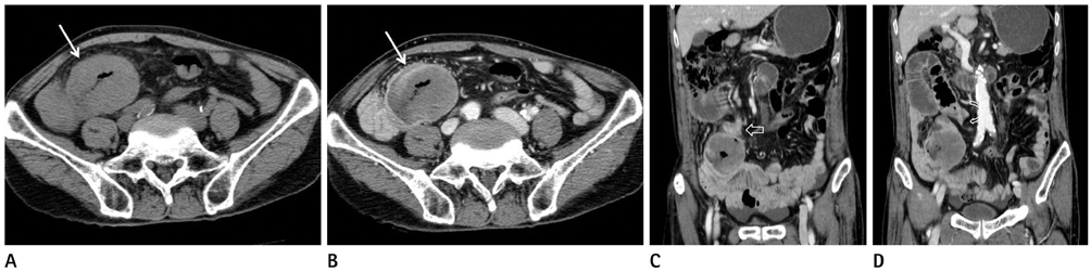
Fig. 1 CT images in a 70-year-old man with abdominal pain. Axial pre-contrast (A) and delayed phase (B) images show a well-defined, round shaped fungating mass (solid arrow) with central necrosis. Coronal reconstruction images on delayed phase CT scan (C, D) show focal wall thickening suggesting tumor seeding (open arrow in C) and beaking appearance (curved arrow in D) with proximal bowel dilatation.
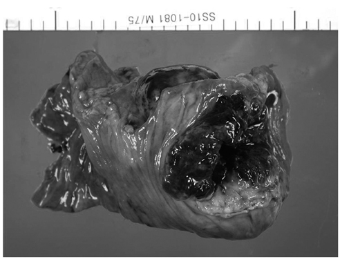
Fig. 2 Photograph of the surgical specimen. The gross specimen shows a fungating necrotic mass in the jejunum.
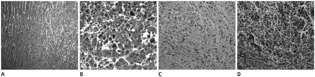
Fig. 3 Pathologic findings of the mass [A. H&E (× 100). B. H&E (× 400). C. Cytokeratin (× 200). D. Vimentin (× 200)]. Microscopically, the tumor consisted mostly of discohesive polygonal giant cells with a few spindle cells (A, B). Tumor cells showed focal strong staining for cytokeratin (C) and diffuse strong staining for vimentin (D).
Reference
-
1. Iezzoni JC, Mills SE. Sarcomatoid carcinomas (carcinosarcomas) of the gastrointestinal tract: a review. Semin Diagn Pathol. 1993; 10:176–187.2. Robey-Cafferty SS, Silva EG, Cleary KR. Anaplastic and sarcomatoid carcinoma of the small intestine: a clinicopathologic study. Hum Pathol. 1989; 20:858–863.3. Padma SK, Permi HS, Patil C, Mathias M. Aggressive monophasic sarcomatoid carcinoma of small intestine - a rare case report with review of literature. NUJHS. 2012; 2:45–47.4. Agrawal S, Trivedi MH, Lukens FJ, Moon C, Ingram EA, Barthel JS. Anaplastic and sarcomatoid carcinoma of the small intestine: an unusual tumor. J Clin Gastroenterol. 1999; 29:99–101.5. Yucel AF, Kocakusak A, Arikan S, Demirbag N, Tarlaci A, Batur S. A rare cause of acute abdomen: perforated primary sarcomatoid carcinoma of the small intestine - report of a case, with a brief review of the literature. J Cancer Res Ther. 2011; 7:348–350.6. Reid-Nicholson M, Idrees M, Perino G, Hytiroglou P. Sarcomatoid carcinoma of the small intestine: a case report and review of the literature. Arch Pathol Lab Med. 2004; 128:918–921.7. Lee SE, Park SY. Sarcomatoid carcinoma of the small intestine: a rare and highly aggressive tumor. J Korean Surg Soc. 2012; 83:321–324.8. Moriwaki Y, Sugiyama M. Severe anemia inducing preshock caused by sarcomatoid carcinoma of the small intestine. Int Surg. 2009; 94:164–170.9. Tsukadaira A, Koizumi T, Okubo Y, Takashi S, Koide N, Arai K, et al. Small-intestinal sarcomatoid carcinoma with superior vena cava syndrome. J Gastroenterol. 2002; 37:471–475.10. Paik HJ, Choi YM. A case of carcinosarcoma in duodenum. J Korean Surg Soc. 1991; 41:549–553.
- Full Text Links
-
- Actions
-
Cited
- CITED
-
- Close
- Share
-
- Similar articles
-
- Sarcomatoid Carcinoma of the Duodenum: A case report
- Sarcomatoid carcinoma of the small intestine: a rare and highly aggressive tumor
- A Surgically Resected Large Sarcomatoid Carcinoma of the Jejunum: A Case Report and Literature Review
- A Case of Vulvar Carcinoma: Squamous Cell Carcinoma with Sarcomatoid Features
- A Case of Sarcomatoid Renal Cell Carcinoma with Multiple Renal Stones and Pyonephrosis
