J Korean Soc Radiol.
2013 Oct;69(4):279-282. 10.3348/jksr.2013.69.4.279.
Anomalous Pulmonary Venous Return: A Case Report
- Affiliations
-
- 1Department of Radiology, Sanggye Paik Hospital, Inje University College of Medicine, Seoul, Korea. s2621@paik.ac.kr
- 2Department of Emergency Medicine, Sanggye Paik Hospital, Inje University College of Medicine, Seoul, Korea.
- KMID: 2041947
- DOI: http://doi.org/10.3348/jksr.2013.69.4.279
Abstract
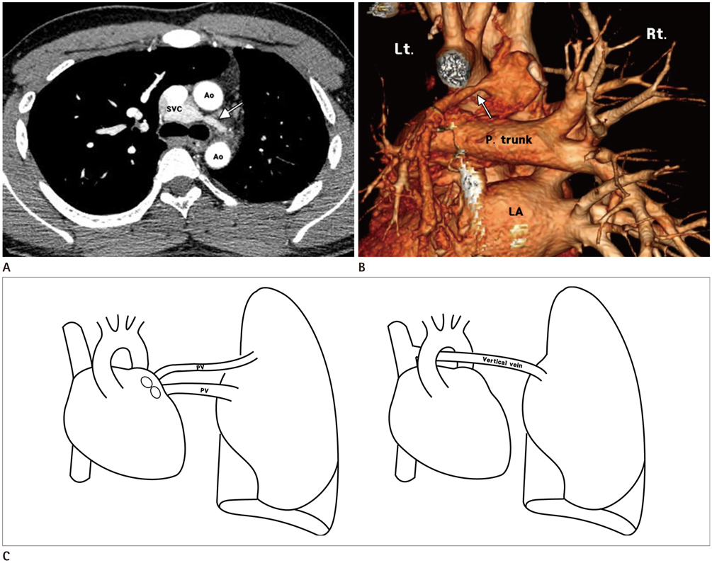
- Partial anomalous pulmonary venous return is a type of congenital pulmonary venous anomaly. We present a rare type of partial pulmonary venous return, subaortic vertical vein drains left lung to superior vena cava, accompanying hypoplasia of the ipsilateral lung and pulmonary artery. We also review the previous report and relationship of these structures.
Figure
Cited by 2 articles
-
The Effect of Two-Injection Ethanol Sclerotherapy with 5-Minute Duration of Exposure Time in Simple Renal Cysts
Dong Eon Kim, Seung Eun Lee, Mi-Jin Kang, Jae Ho Cho, Jihae Lee, Kyung Eun Bae, Jae Hyung Kim, Tae Kyung Kang, Soung Hee Kim, Ji-Young Kim, Myeong Ja Jeong, Soo Hyun Kim
J Korean Soc Radiol. 2017;77(2):113-120. doi: 10.3348/jksr.2017.77.2.113.Anomalous Pulmonary Venous Return Accompanied by Normal Superior Pulmonary Veins in the Left Upper Lobe: A Case Report
Dong Eon Kim, Mi-Jin Kang, Jihae Lee, Kyung Eun Bae, Jae Hyung Kim, Tae Kyung Kang, Soung Hee Kim, Ji-Young Kim, Myeong Ja Jeong, Soo Hyun Kim
J Korean Soc Radiol. 2017;77(2):125-128. doi: 10.3348/jksr.2017.77.2.125.
Reference
-
1. Dillman JR, Yarram SG, Hernandez RJ. Imaging of pulmonary venous developmental anomalies. AJR Am J Roentgenol. 2009; 192:1272–1285.2. Ho ML, Bhalla S, Bierhals A, Gutierrez F. MDCT of partial anomalous pulmonary venous return (PAPVR) in adults. J Thorac Imaging. 2009; 24:89–95.3. Zylak CJ, Eyler WR, Spizarny DL, Stone CH. Developmental lung anomalies in the adult: radiologic-pathologic correlation. Radiographics. 2002; 22 Spec No:S25–S43.4. ElBardissi AW, Dearani JA, Suri RM, Danielson GK. Left-sided partial anomalous pulmonary venous connections. Ann Thorac Surg. 2008; 85:1007–1014.5. Demos TC, Posniak HV, Pierce KL, Olson MC, Muscato M. Venous anomalies of the thorax. AJR Am J Roentgenol. 2004; 182:1139–1150.6. Kurkcuoglu IC, Eroglu A, Karaoglanoglu N, Polat P. Pulmonary hypoplasia in a 52-year-old woman. Ann Thorac Surg. 2005; 79:689–691.7. Fletcher BD, Garcia EJ, Colenda C, Borkat G. Reduced lung volume associated with acquired pulmonary artery obstruction in children. AJR Am J Roentgenol. 1979; 133:47–52.8. Davies G, Reid L. Growth of the alveoli and pulmonary arteries in childhood. Thorax. 1970; 25:669–681.9. Dixit R, Kumar J, Chowdhury V, Rajeshwari K, Sethi GR. Case report: isolated unilateral pulmonary vein atresia diagnosed on 128-slice multidetector CT. Indian J Radiol Imaging. 2011; 21:253–256.10. Lee HN, Kim YT, Cho SS. Individual pulmonary vein atresia in adults: report of two cases. Korean J Radiol. 2011; 12:395–399.11. Beerman LB, Oh KS, Park SC, Freed MD, Sondheimer HM, Fricker FJ, et al. Unilateral pulmonary vein atresia: clinical and radiographic spectrum. Pediatr Cardiol. 1983; 4:105–112.
- Full Text Links
- Actions
-
Cited
- CITED
-
- Close
- Share
- Similar articles
-
- A Case of Surgically Corrected-Combined form of Total Anomalous Pulmonary Venous Return
- Anomalous Pulmonary Venous Return Accompanied by Normal Superior Pulmonary Veins in the Left Upper Lobe: A Case Report
- Levoatriocardinal Vein Combined with Pulmonary Venous Varix Mimicking Arteriovenous Malformations: A Case Report
- Partial Anomalous Pulmonary Venous Return into Coronary Sinus with Intact Atrial Septum
- Congenital Pulmonary Lymphangiectasia, Associated with Total Anomalous Pulmonary Venous Return


