Epithelial-Myoepithelial Carcinoma in Nasal Cavity with Bony Destruction: A Case Report
- Affiliations
-
- 1Department of Radiology, Ilsan Paik Hospital, Inje University School of Medicine, Goyang, Korea. hoonbeer@hanmail.net
- KMID: 2041944
- DOI: http://doi.org/10.3348/jksr.2013.69.4.265
Abstract
- Epithelial-myoepithelial carcinoma (EMC) is a rare tumor that commonly involves the salivary glands. EMC arising from the nasal cavity is one of the most unusual cases. We describe a case of a 48-year-old patient who is presented with bilateral nasal obstruction for several months. Multidetector computed tomography reveals expansile, well-defined, heterogeneous enhancing soft tissue masses filling the nasal cavity with bony destruction of hard palate and maxillary alveolar ridge. The carcinoma was histologically characterized by a mixture of trabecular structure with myoepithelial cells and ductal cells, which are confirmed by electron microscopy and immunohistochemistry.
MeSH Terms
Figure
-
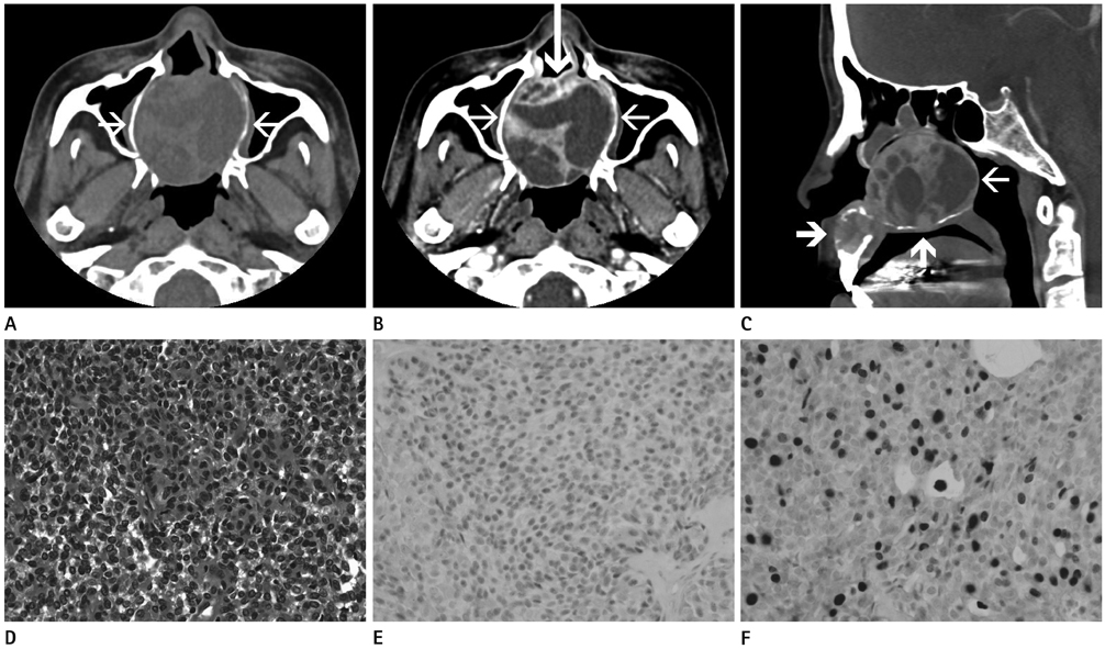
Fig. 1 A 48-year-old woman with epithelial-myoepithelial tumor in nasal cavity. A. An expansile well-marginated soft tissue mass, filling the nasal cavity, shows heterogenous nature on non-enhancing axial CT scan (arrows). B. On enhanced axial CT scan shows multiloculated low attenuation lesions (thin arrows) and septum like enhancing portion in mass (thick arrow). C. Mass extends into nasopharyngeal space posteriorly (thin arrow) and there was also bony destruction of hard palate and maxillary alveolar ridge due to tumor invasion on sagittal CT sacn (thick arrows). D. Each tubules are lined by two layers of cells, inner eosinophilic epithelial cells and outer myoepithelial cells (hematoxylin-eosin, × 400). E. Immunohistochemical analysis shows that the expression of p63 (original magnification, × 400). F. Immunohistochemical analysis shows that the expression of Ki67 (original magnification, × 400).
Reference
-
1. Ellis GI, Auclair PL. Tumors of the Salivary Glands. In : Rosai J, editor. Malignant epithelial tumors. Washington, DC: Armed Forces Institute of Pathology;1996. p. 268–289.2. Batsakis JG, el-Naggar AK, Luna MA. Epithelial-myoepithelial carcinoma of salivary glands. Ann Otol Rhinol Laryngol. 1992; 101:540–542.3. Lee HM, Kim AR, Lee SH. Epithelial-myoepithelial carcinoma of the nasal cavity. Eur Arch Otorhinolaryngol. 2000; 257:376–378.4. M'sakni I, Laabidi B, Bougrine F, Sabbegh-Znaïdi N, Benzarti S, Chebbi K, et al. [Epithelial-myoepithelial carcinoma of the nasal cavity]. Ann Otolaryngol Chir Cervicofac. 2007; 124:228–231.5. Yamanegi K, Uwa N, Hirokawa M, Ohyama H, Hata M, Yamada N, et al. Epithelial-myoepithelial carcinoma arising in the nasal cavity. Auris Nasus Larynx. 2008; 35:408–413.6. Kim HS, Lee WM, Choi SM. Myoepitheliomas of the soft palate: helical CT findings in two patients. Korean J Radiol. 2007; 8:552–555.7. Harada H, Kashiwagi SI, Fujiura H, Kusukawa J, Morimatsu M. Epithelial-myoepithelial carcinoma--report of a case arising in the nasal cavity. J Laryngol Otol. 1996; 110:397–400.8. Jin XL, Ding CN, Chu Q. Epithelial-myoepithelial carcinoma arising in the nasal cavity: a case report and review of literature. Pathology. 1999; 31:148–151.9. Park JO, Jung CK, Sun DI, Kim MS. An unusual presentation of aggressive epithelial-myoepithelial carcinoma of the nasal cavity with high-grade histology. J Laryngol Otol. 2011; 125:1286–1289.10. Cho KS, Shin SC, Mun MJ, Roh HJ. A case of myoepithelial carcinoma originated from inferior turbinate. Korean J Otorhinolaryngol-Head Neck Surg. 2010; 53:791–794.11. Lee HM, Choi CS, Kim A, Lee SH. Epithelial-myoepithelial carcinoma arising in the nasal cavity-immunohistochemical and electron microscopic study. Korean J Otolaryngol-Head Neck Surg. 2000; 43:383–386.12. Cho SH, Kim HT, Kim MS, Sun DI, Koo Y. A clinical study of malignant neoplasms of the nasal septum. Korean J Otolaryngol-Head Neck Surg. 1998; 41:68–72.13. Compagno J, Wong RT. Intranasal mixed tumors (pleomorphic adenomas): a clinicopathologic study of 40 cases. Am J Clin Pathol. 1977; 68:213–218.14. Spreer J, Krahe T, Jung G, Lackner K. Spiral versus conventional CT in routine examinations of the neck. J Comput Assist Tomogr. 1995; 19:905–910.
- Full Text Links
-
- Actions
-
Cited
- CITED
-
- Close
- Share
-
- Similar articles
-
- Epithelial-myoepithelial Carcinoma Arising in the Nasal Cavity-Immunohistochemical and Electron Microscopic Study
- A Case of Myoepithelial Carcinoma Originated from Inferior Turbinate
- Epithelial-Myoepithelial Carcinoma of the Inferior Turbinate: A Case Report
- Epithelial-myoepithelial carcinoma arising in pleomorphic adenoma of palate
- Epithelial-Myoepithelial Carcinoma of Intercalated Duct of Parotid Gland
