J Korean Soc Radiol.
2011 May;64(5):515-518. 10.3348/jksr.2011.64.5.515.
Traumatic Neuroma in a Breast Cancer Patient After Modified Radical Mastectomy: A Case Report
- Affiliations
-
- 1Department of Radiology, Ajou University School of Medicine, Korea. medhand@ajou.ac.kr
- 2Department of Surgery, Ajou University School of Medicine, Korea.
- 3Department of Pathology, Ajou University School of Medicine, Korea.
- KMID: 2040822
- DOI: http://doi.org/10.3348/jksr.2011.64.5.515
Abstract
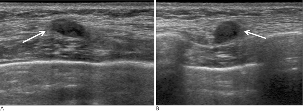
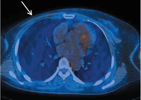
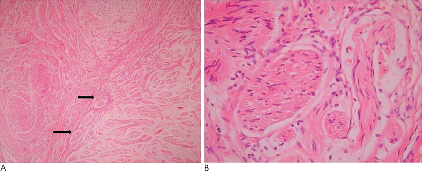
- Traumatic neuromas are rare benign lesions that develop from non-neoplastic proliferation of axons, schwann cells, and fibroblasts at the proximal end of transected or injured nerves as a result of trauma or surgery. We present the case of a traumatic neuroma in a 47-year-old female who was treated with a right modified radical mastectomy for breast cancer 14 years ago. Ultrasound examination revealed an oval-shaped hypoechoic nodule at the 9-o'clock position in the right chest wall. Color Doppler imaging showed no increased blood flow and a positron emission tomography-computed tomography examination revealed no fluorodeoxyglucose uptake in this nodule. The typical histologic findings were present.
MeSH Terms
Figure
Reference
-
1. Foltan R, Klima K, Spackova J, Sedy J. Mechanism of traumatic neuroma development. Med Hypotheses. 2008; 71:572–576.2. Murphey MD, Smith WS, Smith SE, Kransdorf MJ, Temple HT. From the archives of the AFIP. Imaging of musculoskeletal neurogenic tumors: radiologic-pathologic correlation. Radiographics. 1999; 19:1253–1280.3. Rosso R, Scelsi M, Carnevali L. Granular cell traumatic neuroma: a lesion occurring in mastectomy scars. Arch Pathol Lab Med. 2000; 124:709–711.4. Baltalarli B, Demirkan N, Yagci B. Traumatic neuroma: unusual benign lesion occurring in the mastectomy scar. Clin Oncol (R Coll Radiol). 2004; 16:503–504.5. Wang X, Cao X, Ning L. Traumatic neuromas after mastectomy. ANZ J Surg. 2007; 77:704–705.6. Wong L. Intercostal neuromas: a treatable cause of postoperative breast surgery pain. Ann Plast Surg. 2001; 46:481–484.7. Provencher MT, Handfield K, Boniquit NT, Reiff SN, Sekiya JK, Romeo AA. Injuries to the pectoralis major muscle: diagnosis and management. Am J Sports Med. 2010; 38:1693–1705.8. Moosman DA. Anatomy of the pectoral nerves and their preservation in modified mastectomy. Am J Surg. 1980; 139:883–886.9. Yilmaz MH, Esen G, Ayarcan Y, Aydogan F, Ozguroglu M, Demir G, et al. The role of US and MR imaging in detecting local chest wall tumor recurrence after mastectomy. Diagn Interv Radiol. 2007; 13:13–18.10. Beggs I. Sonographic appearances of nerve tumors. J Clin Ultrasound. 1999; 27:363–368.
- Full Text Links
- Actions
-
Cited
- CITED
-
- Close
- Share
- Similar articles
-
- A Traumatic Neuroma in Breast Cancer Patient after Mastectomy: A Case Report
- Breast Cancer During Pregnancy
- Comparison of Psychiatric Symptoms between Total Mastectomy and Breast Conserving Surgery in Breast Cancer Patients
- Breast Cancer during Pregnancy
- Cases with Endometrial Polyp and Endocervical Polyp Associated With Tamoxifen Use


