J Korean Soc Radiol.
2011 May;64(5):509-513. 10.3348/jksr.2011.64.5.509.
Aggressive Behavior of a Giant Cell Tumor Involving the Metacarpal Bone During Pregnancy: Case Report
- Affiliations
-
- 1Department of Radiology, Kyung Hee University Medical Center, Kyung Hee University, Korea. gdluck@hitel.net
- 2Department of Radiology, Kyung Hee University at Gandong, Kyung Hee University, Korea.
- 3Department of Pathology, Kyung Hee University Medical Center, Kyung Hee University, Korea.
- 4Department of Orthopedic Surgery, Kyung Hee University Medical Center, Kyung Hee University, Korea.
- KMID: 2040821
- DOI: http://doi.org/10.3348/jksr.2011.64.5.509
Abstract
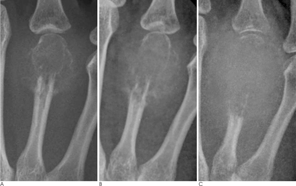
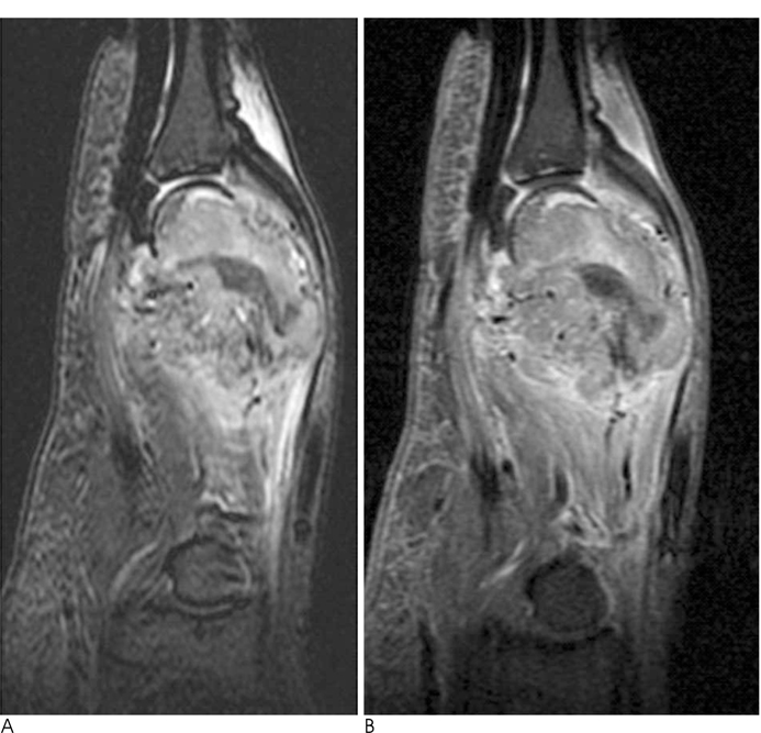
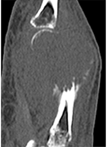
- Giant cell tumors are benign osteolytic tumors with a variable degree of aggressiveness. We report a rare case of a giant cell tumor involving the metacarpal bone, which was detected during pregnancy and showed rapid progression on a follow-up examination.
Figure
Reference
-
1. Murphey MD, Nomikos GC, Flemming DJ, Gannon FH, Temple HT, Kransdorf MJ. From the archives of afip. Imaging of giant cell tumor and giant cell reparative granuloma of bone: radiologicpathologic correlation. Radiographics. 2001; 21:1283–1309.2. James SL, Davies AM. Giant-cell tumours of bone of the hand and wrist: a review of imaging findings and differential diagnoses. Eur Radiol. 2005; 15:1855–1866.3. Turcotte RE, Sim FH, Unni KK. Giant cell tumor of the sacrum. Clin Orthop Relat Res. 1993; 215–221.4. Komiya S, Zenmyo M, Inoue A. Bone tumors in the pelvis presenting growth during pregnancy. Arch Orthop Trauma Surg. 1999; 119:22–29.5. Minhas MS, Mehboob G, Ansari I. Giant cell tumours in hand bones. J Coll Physicians Surg Pak. 2010; 20:460–463.6. Kotnis NA, Davies AM, Kindblom LG, James SL. Giant cell tumour of the triquetrum. Skeletal Radiol. 2009; 38:593–595.7. Maxwell C, Barzilay B, Shah V, Wunder JS, Bell R, Farine D. Maternal and neonatal outcomes in pregnancies complicated by bone and soft-tissue tumors. Obstet Gynecol. 2004; 104:344–348.8. Ross AE, Bojescul JA, Kuklo TR. Giant cell tumor: a case report of recurrence during pregnancy. Spine. 2005; 30:E332–E335.9. Simon MA, Phillips WA, Bonfiglio M. Pregnancy and aggressive or malignant primary bone tumors. Cancer. 1984; 53:2564–2569.10. Ishibe M, Ishibe Y, Ishibashi T, Nojima T, Rosier RN, Puzas JE, et al. Low content of estrogen receptors in human giant cell tumors of bone. Arch Orthop Trauma Surg. 1994; 113:106–109.


