J Korean Surg Soc.
2010 Dec;79(Suppl 1):S67-S70. 10.4174/jkss.2010.79.Suppl1.S67.
Tailgut Cyst in a Neonate
- Affiliations
-
- 1Department of Surgery, College of Medicine, Yeungnam University, Daegu, Korea. nhlee@med.yu.ac.kr
- KMID: 2040557
- DOI: http://doi.org/10.4174/jkss.2010.79.Suppl1.S67
Abstract
- Tailgut cysts are rare congenital lesions thought to arise from vestiges of the tailgut/postanal gut, which is a primitive gut temporarily present at the most caudal portion of the embryo. The lesions are usually multilocular cysts lined by various types of epithelium in the retrorectal space. We experienced a tailgut cyst in an 18-day-old girl initially presented as an epidermoid cyst like skin lesion in the coccygeal region. MRI showed a well-defined multiloculated cystic mass between the rectum and coccyx, measuring 12x23 mm and markedly hyperintense on T2 weighted images and hypointense on T1-weighted images. The cyst was surgically removed via a posterior sagittal approach and microscopically lined by ciliated columnar, transitional and squamous cells. Tailgut cysts are usually found in adult females, and extremely rarely in neonates. We report a tailgut cyst in a neonate with a review of the literature.
Keyword
MeSH Terms
Figure
-
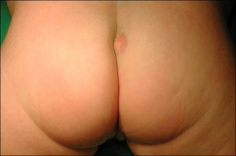
Fig. 1 Photograph shows a subcutaneous epidermoid cyst-like lesion in the coccygeal region.

Fig. 2 Ultrasonography shows a multiseptated cystic mass abutting the coccyx.
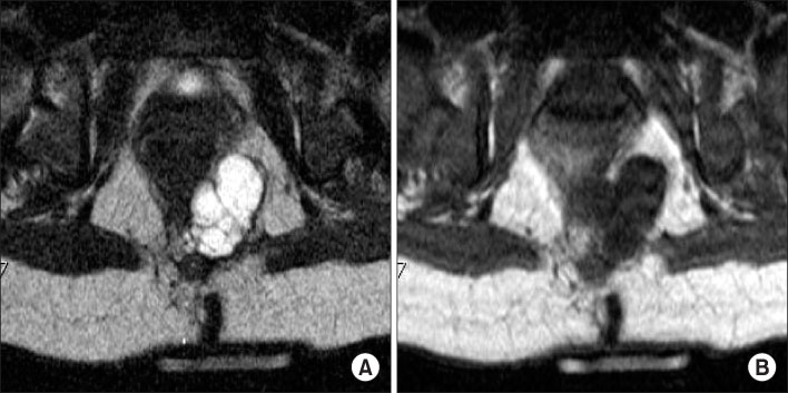
Fig. 3 Axial MR images show a multiloculated cystic mass in the retrorectal space, which is markedly hyperintense on T2-weighted images (A), and hypointense on T1-weighted images (B), respectively.

Fig. 4 The microscopic pathologic finding shows multiloculated cysts, which are lined by ciliated columnar, transitional and squamous cells (H&E stain, ×100).
Reference
-
1. Hjermstad BM, Helwig EB. Tailgut cysts. Report of 53 cases. Am J Clin Pathol. 1988. 89:139–147.2. Killingsworth C, Gadacz TR. Tailgut cyst (retrorectal cystic hamartoma): report of a case and review of the literature. Am Surg. 2005. 71:666–673.3. Oh JT, Son SW, Kim MJ, Kim L, Kim H, Hwang EH. Tailgut cyst in a neonate. J Pediatr Surg. 2000. 35:1833–1835.4. Nakagawa M, Hara M, Oshima H, Kitase M, Shibamoto Y. Radiological findings of tailgut cyst in a fetus. J Comput Assist Tomogr. 2008. 32:210–213.5. Kanthan SC, Kanthan R. Unusual retrorectal lesion. Asian J Surg. 2004. 27:144–146.6. Gönül II, Bağlan T, Pala I, Menteş B. Tailgut cysts: diagnostic challenge for both pathologists and clinicians. Int J Colorectal Dis. 2007. 22:1283–1285.7. Prasad AR, Amin MB, Randolph TL, Lee CS, Ma CK. Retrorectal cystic hamartoma: report of 5 cases with malignancy arising in 2. Arch Pathol Lab Med. 2000. 124:725–729.8. Au E, Anderson O, Morgan B, Alarcon L, George ML. Tailgut cysts: report of two cases. Int J Colorectal Dis. 2009. 24:345–350.9. Aflalo-Hazan V, Rousset P, Mourra N, Lewin M, Azizi L, Hoeffel C. Tailgut cysts: MRI findings. Eur Radiol. 2008. 18:2586–2593.10. Hall DA, Pu RT, Pang Y. Diagnosis of foregut and tailgut cysts by endosonographically guided fine-needle aspiration. Diagn Cytopathol. 2007. 35:43–46.
