Korean J Obstet Gynecol.
2010 Dec;53(12):1141-1145. 10.5468/kjog.2010.53.12.1141.
Ectopic ovary with a mature cystic teratoma diagnosed by laparoscopy: A case report
- Affiliations
-
- 1Department of Obstetrics and Gynecology, The Catholic University of Korea College of Medicine, Seoul, Korea. mrkim@catholic.ac.kr
- KMID: 2037517
- DOI: http://doi.org/10.5468/kjog.2010.53.12.1141
Abstract
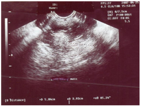
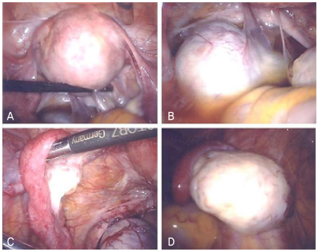
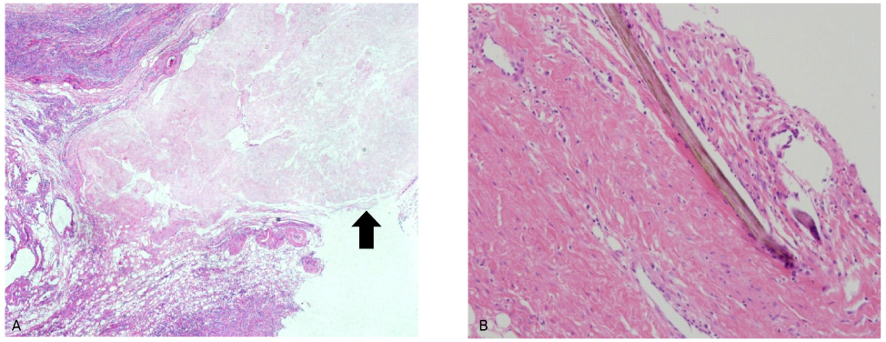
- The ectopic ovary is a rarely reported gynecologic entity. A variety of synonymous terms have been used to describe this condition, such as supernumerary ovary, accessory ovary, and ovarian implant syndrome. The etiology of ectopic ovary is poorly understood. The ectopic ovaries may occur in two ways. First, in the embryonic theories, they are believed to result from abnormal separation of a small portion of the developing and migrating ovarian primordium. Second, the accessory ovary can occur from acquired conditions such as inflammation and operations. In this report, we describe a case of the ectopic ovary with a mature cystic teratoma autoamputated into the cul-de-sac and subsequently diagnosed by laparoscopy.
Keyword
MeSH Terms
Figure
Reference
-
1. Kusaka M, Mikuni M. Ectopic ovary: a case of autoamputated ovary with mature cystic teratoma into the cul-de-sac. J Obstet Gynaecol Res. 2007. 33:368–370.2. Lachman MF, Berman MM. The ectopic ovary. A case report and review of the literature. Arch Pathol Lab Med. 1991. 115:233–235.3. Kuga T, Esato K, Takeda K, Sase M, Hoshii Y. A supernumerary ovary of the omentum with cystic change: report of two cases and review of the literature. Pathol Int. 1999. 49:566–570.4. Ushakov FB, Meirow D, Prus D, Libson E, BenShushan A, Rojansky N. Parasitic ovarian dermoid tumor of the omentum-A review of the literature and report of two new cases. Eur J Obstet Gynecol Reprod Biol. 1998. 81:77–82.5. Wharton LR. Two cases of supernumerary ovary and one of accessory ovary, with an analysis of previously reported cases. Am J Obstet Gynecol. 1959. 78:1101–1119.6. Lim MC, Park SJ, Kim SW, Lee BY, Lim JW, Lee JH, et al. Two dermoid cysts developing in an accessory ovary and an eutopic ovary. J Korean Med Sci. 2004. 19:474–476.7. Levy B, DeFranco J, Parra R, Holtz P. Intrarenal supernumerary ovary. J Urol. 1997. 157:2240–2241.8. Moon W, Kim Y, Rhim H, Koh B, Cho O. Coexistent cystic teratoma of the omentum and ovary: report of two cases. Abdom Imaging. 1997. 22:516–518.
- Full Text Links
- Actions
-
Cited
- CITED
-
- Close
- Share
- Similar articles
-
- A Case of Papillary Carcinoma of Thyroid Gland Arising from Ovarian Mature Cystic Teratoma
- MR Findings of Malignant Struma Ovarii Associated with Mature Cystic Teratoma of Contralateral Ovary: Case Report
- A Case of Delayed Operated Huge Mature Cystic Teratoma of the Ovary
- Traumatic Rupture of a Mature Cystic Teratoma of the Ovary
- A Case of Adenocarcinoma Arising in Mature Cystic Teratoma of the Ovary


