What is the effect of initial implant position on the crestal bone level in flap and flapless technique during healing period?
- Affiliations
-
- 1Department of Oral Surgery, MAHSA Dental College, Kuala Lumpur, Malaysia. doctor_mohamed_2006@yahoo.com
- 2Department of Oral and Maxillofacial Surgery, Universiti Sains Malaysia School of Dental Sciences, Kubang Kerian, Malaysia.
- 3Department of Periodotology, Universiti Sains Malaysia School of Dental Sciences, Kubang Kerian, Malaysia.
- 4Department of Conservative, MAHSA Dental College, Kuala Lumpur, Malaysia.
- 5Department of Prosthodontics, MAHSA Dental College, Kuala Lumpur, Malaysia.
- KMID: 2027810
- DOI: http://doi.org/10.5051/jpis.2013.43.4.153
Abstract
- PURPOSE
The level of the implant above the marginal bone and flap design have an effect on the bone resorption during the healing period. The aim of this study is to detect the relationship between the level of the implant at the implant placement and the bone level at the healing period in the mesial and distal side of implants placed with flapless (FL) and full-thickness flap (FT) methods.
METHODS
Twenty-two nonsubmerged implants were placed with the FL and FT technique. Periapical radiographs were taken of the patient at implant placement, and at 6 and 12 weeks. By using computer software, bone level measurements were taken from the shoulder of the healing cap to the first bone implant contact in the mesial and distal side of the implant surface.
RESULTS
At 6 weeks, the correlation between the crestal bone level at the implant placement and crestal bone level of the FT mesially was significant (Pearson correlation coefficient=0.675, P<0.023). At 12 weeks, in the FT mesially, the correlation was nonsignificant (Spearman correlation coefficient=0.297, P<0.346). At 6 weeks in the FT distally, the correlation was nonsignificant (Pearson correlation coefficient=0.512, P<0.107). At 12 weeks in the FT distally, the correlation was significant (Spearman correlation coefficient=0.730, P<0.011). At 6 weeks in the FL mesially, the correlation was nonsignificant (Spearman correlation coefficient=0.083, P<0.809). At 12 weeks in the FL mesially, the correlation was nonsignificant (Spearman correlation coefficient=0.062, P<0.856). At 6 weeks in the FL distally, the correlation was nonsignificant (Spearman correlation coefficient=0.197, P<0.562). At 12 weeks in the FL distally, the correlation was significant (Pearson correlation coefficient=0.692, P<0.018).
CONCLUSIONS
A larger sample size is recommended to verify the conclusions in this preliminary study. The bone level during the healing period in the FT was more positively correlated with the implant level at implant placement than in the FL.
Figure
-
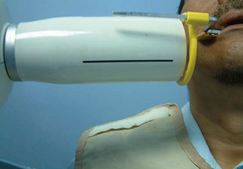
Figure 1 Using the long-cone parallel technique with a Rinn film holder (Dentsply International Inc.).

Figure 2 Correlation between the mesial crestal bone level of the full-thickness flap (FT) at implant placement and the mesial crestal bone level of the FT at 6 weeks (mm). There was a significant positive correlation (moderate to good correlation) (r=0.675, P<0.023).

Figure 3 Correlation between the mesial crestal bone level of the full-thickness flap (FT) at implant placement and the mesial crestal bone level of the FT at 12 weeks (mm). There was a fair statistically nonsignificant positive correlation (r=0. 297, P<0.346).
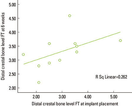
Figure 4 Correlation between the distal crestal bone level of the full-thickness flap (FT) at implant placement and the distal crestal bone level of the FT at 6 weeks (mm). There was a nonsignificant positive correlation (moderate correlation) (r=0. 512, P<0.107).
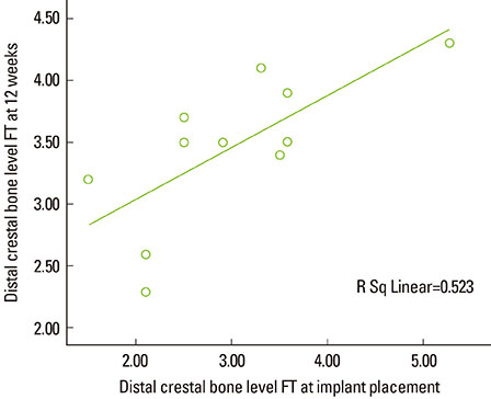
Figure 5 Correlation between the distal crestal bone level of the full-thickness flap (FT) at implant placement and the distal crestal bone level of the FT group at 12 weeks (mm). There was a significant positive correlation (good correlation) (r=0.730, P<0.011).

Figure 6 Correlation between the mesial crestal bone level of the flapless (FL) at implant placement and the mesial crestal bone level of the FL at 6 weeks (mm). There was a nonsignificant positive correlation (poor correlation) (r=0.083, P<0.809).
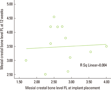
Figure 7 Correlation between the mesial crestal bone level of the flapless (FL) at implant placement and the mesial crestal bone level of the FL at 12 weeks (mm). There was a non-significant positive correlation (poor correlation) (r=0.062, P<0.856).
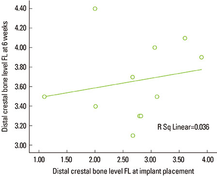
Figure 8 Correlation between the distal crestal bone level of the flapless (FL) at implant placement and the distal crestal bone level of the FL at 6 weeks (mm). There was a poor nonsignificant positive correlation (r=0.197, P<0.562).

Figure 9 Correlation between the distal crestal bone level of the flapless (FL) at implant placement and the distal crestal bone level of the FL at 12 weeks (mm). There was a good statistically significant positive correlation (r=0.692, P<0.018).
Reference
-
1. Bergkvist G, Sahlholm S, Nilner K, Lindh C. Implant-supported fixed prostheses in the edentulous maxilla. A 2-year clinical and radiological follow-up of treatment with non-submerged ITI implants. Clin Oral Implants Res. 2004; 15:351–359.
Article2. Bischof M, Nedir R, Abi Najm S, Szmukler-Moncler S, Samson J. A five-year life-table analysis on wide neck ITI implants with prosthetic evaluation and radiographic analysis: results from a private practice. Clin Oral Implants Res. 2006; 17:512–520.
Article3. Soadoun AP, Touati B. Soft tissue recession around implants: is it still unavoidable?--Part II. Pract Proced Aesthet Dent. 2007; 19:81–87.4. Brägger U, Hafeli U, Huber B, Hammerle CH, Lang NP. Evaluation of postsurgical crestal bone levels adjacent to non-submerged dental implants. Clin Oral Implants Res. 1998; 9:218–224.
Article5. Oh TJ, Shotwell JL, Billy EJ, Wang HL. Effect of flapless implant surgery on soft tissue profile: a randomized controlled clinical trial. J Periodontol. 2006; 77:874–882.
Article6. Ostman PO, Hellman M, Albrektsson T, Sennerby L. Direct loading of Nobel Direct and Nobel Perfect one-piece implants: a 1-year prospective clinical and radiographic study. Clin Oral Implants Res. 2007; 18:409–418.
Article7. Lee DW, Choi YS, Park KH, Kim CS, Moon IS. Effect of microthread on the maintenance of marginal bone level: a 3-year prospective study. Clin Oral Implants Res. 2007; 18:465–470.
Article8. Hartman GA, Cochran DL. Initial implant position determines the magnitude of crestal bone remodeling. J Periodontol. 2004; 75:572–577.
Article9. Novaes AB Jr, de Oliveira RR, Muglia VA, Papalexiou V, Taba M. The effects of interimplant distances on papilla formation and crestal resorption in implants with a morse cone connection and a platform switch: a histomorphometric study in dogs. J Periodontol. 2006; 77:1839–1849.
Article10. Cochran DL, Hermann JS, Schenk RK, Higginbottom FL, Buser D. Biologic width around titanium implants: a histometric analysis of the implanto-gingival junction around unloaded and loaded nonsubmerged implants in the canine mandible. J Periodontol. 1997; 68:186–198.
Article11. Hürzeler M, Fickl S, Zuhr O, Wachtel HC. Peri-implant bone level around implants with platform-switched abutments: preliminary data from a prospective study. J Oral Maxillofac Surg. 2007; 65:7 Suppl 1. 33–39.
Article12. Choi BH, Li J, Kim HS, Ko CY, Jeong SM, Xuan F. Comparison of submerged and nonsubmerged implants placed without flap reflection in the canine mandible. Oral Surg Oral Med Oral Pathol Oral Radiol Endod. 2008; 105:561–565.
Article13. Machtei EE, Oved-Peleg E, Peled M. Comparison of clinical, radiographic and immunological parameters of teeth and different dental implant platforms. Clin Oral Implants Res. 2006; 17:658–665.
Article14. Papalexiou V, Novaes AB Jr, Ribeiro RF, Muglia V, Oliveira RR. Influence of the interimplant distance on crestal bone resorption and bone density: a histomorphometric study in dogs. J Periodontol. 2006; 77:614–621.
Article15. Jeong SM, Choi BH, Li J, Kim HS, Ko CY, Jung JH, et al. Flapless implant surgery: an experimental study. Oral Surg Oral Med Oral Pathol Oral Radiol Endod. 2007; 104:24–28.
Article16. Gomez-Roman G. Influence of flap design on peri-implant interproximal crestal bone loss around single-tooth implants. Int J Oral Maxillofac Implants. 2001; 16:61–67.17. Ozan O, Turkyilmaz I, Yilmaz B. A preliminary report of patients treated with early loaded implants using computerized tomography-guided surgical stents: flapless versus conventional flapped surgery. J Oral Rehabil. 2007; 34:835–840.
Article18. al-Ansari BH, Morris RR. Placement of dental implants without flap surgery: a clinical report. Int J Oral Maxillofac Implants. 1998; 13:861–865.19. Auty C, Siddiqui A. Punch technique for preservation of interdental papillae at nonsubmerged implant placement. Implant Dent. 1999; 8:160–166.
Article20. Gibney JW. Minimally invasive implant surgery. J Oral Implantol. 2001; 27:73–76.
Article21. Marchack CB. CAD/CAM-guided implant surgery and fabrication of an immediately loaded prosthesis for a partially edentulous patient. J Prosthet Dent. 2007; 97:389–394.
Article22. Al-Juboori MJ, Bin Abdulrahaman S, Jassan A. Comparison of flapless and conventional flap and the effect on crestal bone resorption during a 12-week healing period. Dent Implantol Update. 2012; 23:9–16.23. Al-Juboori MJ, bin Abdulrahaman S, Subramaniam R, Tawfiq OF. Less morbidity with flapless implant. Dent Implantol Update. 2012; 23:25–30.24. Todescan FF, Pustiglioni FE, Imbronito AV, Albrektsson T, Gioso M. Influence of the microgap in the peri-implant hard and soft tissues: a histomorphometric study in dogs. Int J Oral Maxillofac Implants. 2002; 17:467–472.25. Hermann JS, Schoolfield JD, Nummikoski PV, Buser D, Schenk RK, Cochran DL. Crestal bone changes around titanium implants: a methodologic study comparing linear radiographic with histometric measurements. Int J Oral Maxillofac Implants. 2001; 16:475–485.26. Hermann JS, Buser D, Schenk RK, Higginbottom FL, Cochran DL. Biologic width around titanium implants. A physiologically formed and stable dimension over time. Clin Oral Implants Res. 2000; 11:1–11.
Article27. Tarnow DP, Cho SC, Wallace SS. The effect of inter-implant distance on the height of inter-implant bone crest. J Periodontol. 2000; 71:546–549.
Article28. Rao W, Benzi R. Single mandibular first molar implants with flapless guided surgery and immediate function: preliminary clinical and radiographic results of a prospective study. J Prosthet Dent. 2007; 97:6 Suppl. S3–S14.
Article29. Wyatt CC, Zarb GA. Bone level changes proximal to oral implants supporting fixed partial prostheses. Clin Oral Implants Res. 2002; 13:162–168.
Article
- Full Text Links
-
- Actions
-
Cited
- CITED
-
- Close
- Share
-
- Similar articles
-
- Blood Vessels of the Peri-Implant Mucosa: a Comparison Between Flap and Flapless Procedures
- The effects of tissue punch diameter on healing around implants in flapless implant surgery
- Radiographic evaluation of marginal bone level alteration around narrow implants placed in narrow alveolar ridge using guided flapless surgery
- Radiographic evaluation of the proximal bone level between two implants: A 3-year comparative study between Branemark and ITI implants in the mandibular posterior region
- Installation Immediate Implant with Sinus Lift Crestal Approach Technique: A Case Report of 4-year Follow-up
