Spinal Arachnoid Cyst: Treated with Pars Osteotomy and Recapping Laminoplasty: Report of 5 Cases
- Affiliations
-
- 1Spine Center, Korea Hospital, Pusan, Korea.
- 2Department of Orthopaedic Surgery, Daedong Hospital, Pusan, Korea. ahnsjosdept@naver.com
- KMID: 2003117
- DOI: http://doi.org/10.4184/jkss.2009.16.3.215
Abstract
- Spinal arachnoid cysts are a rare disease with an unknown origin. Because of their broad base, a total laminectomy with or without fusion has been the treatment of the choice. We encountered 5 patients with a spinal arachnoid cyst who were treated by recapping laminoplasty after pars osteotomy. This procedure has not been reported in Korea. All patients showed neurological recovery with no recurrence of the cyst. The findings on the stressed plain film confirmed bony union and stability of the posterior element.
Figure
-

Fig. 1. (A) Preoperative MRI and plain X-ray shows type III arachoid cyst from L2 to L5 in the 43-year-old female patient. (B) Intraoperative photograph. we try to remain distal supraspinous ligament attached distal part and complete removal of cyst 9cm in length. (C) Postoperative-6-month X-ray shows good union of pars after recapping and interosseous wiring and pars fixation using screw.
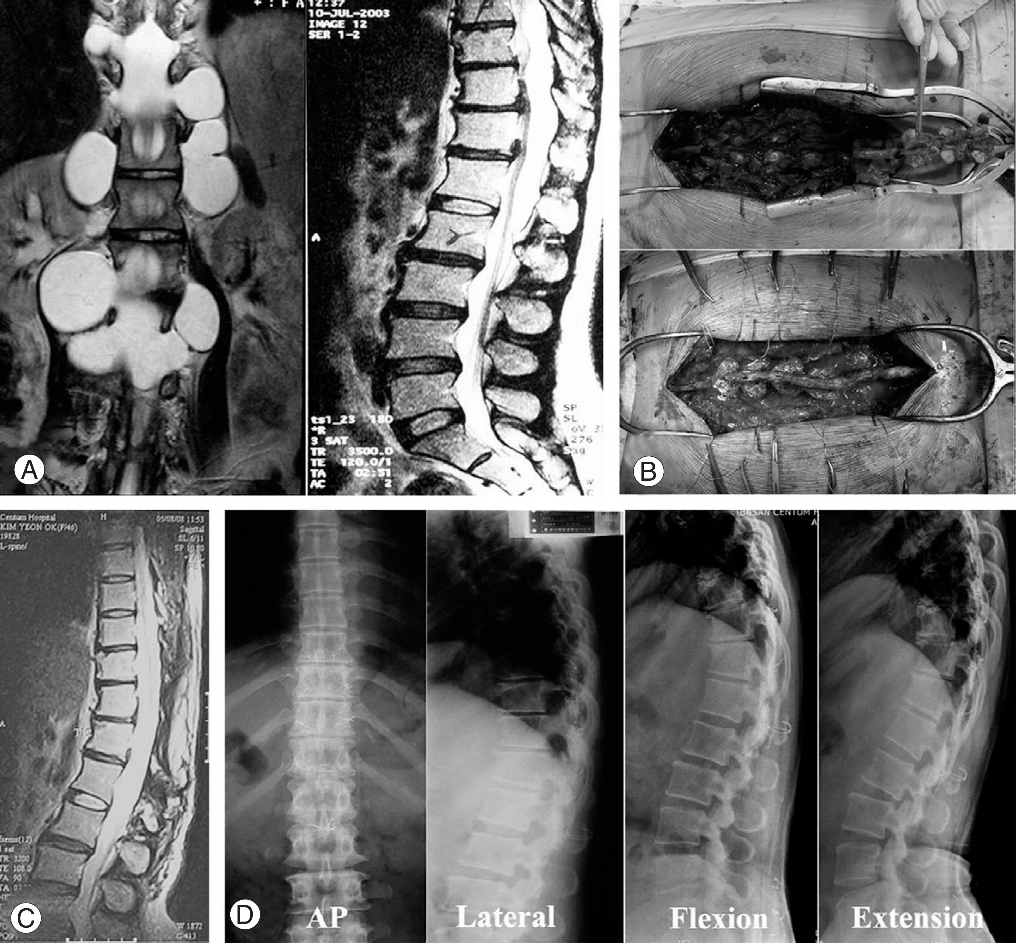
Fig. 2. (A) Preoperative MRI shows type I arachnoid cyst from T9 to L2 in the 44-year-old female patient. (B) Intraoperative photograph shows remaing distal supraspinous ligament and wire fixation. (C) Postoperative MRI and X-ray shows complete removal of cyst. (D) Postoperative 3 year after recapping laminoplasty shows complete union at the osteotmy levels and shows no instability.
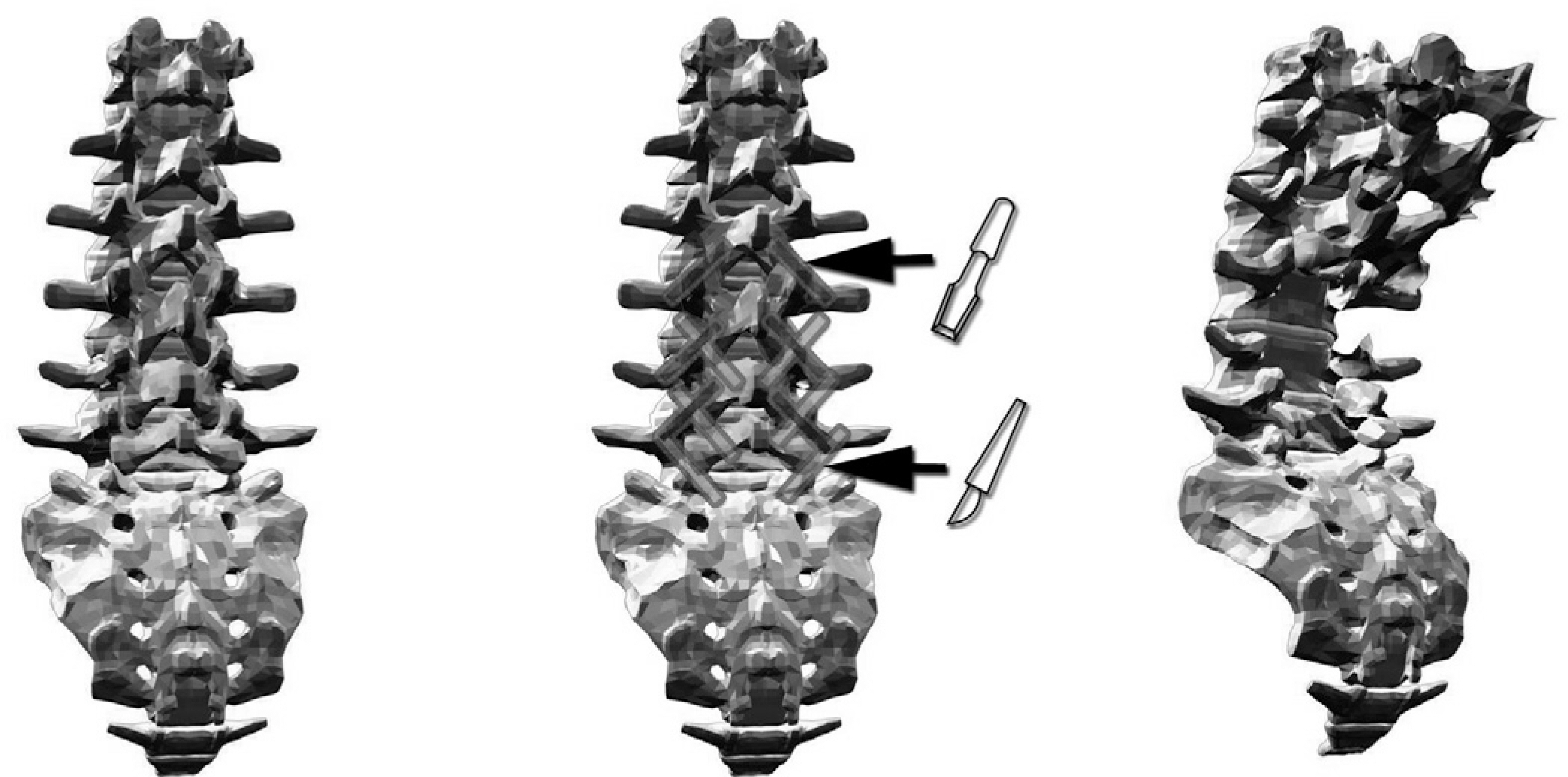
Fig. 3. Schematic diagram of recapping laminoplasty.
Reference
-
1). Dastur HM. The radiological appearances of spinal extradural arachnoid cysts. J Neurol Neurosurg Psychiar-ty. 1963; 26:231–235.
Article2). Rabb CH, McComb JG, Raffel C, Kennedy JG. Spinal arachnoid cysts in the pediatric age group: an association with neural tube defects. J Neurosurg. 1992; 77:369–372.
Article3). Rimmelin A, Clouet PL, Salatino S, et al. .:. Imaging of thoracic and lumbar spinal extradural arachnoid cysts: report of two cases. Neuroradiology. 1997; 39:203–206.
Article4). Kulkarin AG, Goel A, Thiruppathy SP, Desai K. Extradural arachnoid cysts: a study of seven cases. British Journal of Neurology. 2004; 18:484–488.5). Krings T, Lukas R, Reul J, et al. .:. Diagnostic and Therapeutic management of spinal arachnoid cysts. Acta Neurochir. 2001; 143:227–235.
Article6). Choi JY, Kim SH, Lee WS, Sung KH. Spinal extradural arachnoid cyst. Acta Neurochir. 2006; 148:579–585.
Article7). Doita M, Nishida K, Miura J, Takada T, Kurosaka M, Fujii M. Kinematic magnetic resonance imaging of a thoracic spinal extradural arachnoid cyst: an alternative suggestion for exacerbation of symptoms during straining. Spine. 2003; 28:229–233.
Article8). Cloward RB. Congenital spinal extradural cyst: case report with review of literiture. Ann Surg. 1968; 168:851–864.9). Hatashita S, Kondo A, Shimizu T, Kurosu A, Ueno H. Spinal extradural arachnoid cyst. Case report. Neurol Med Chir (Tokyo). 2001; 41:318–321.
Article10). Kawahara N, Tomita K, Shinya Y, et al. .:. Recapping T-saw laminoplasty for spinal cord tumors. 1999; 24:1363–1370.
- Full Text Links
-
- Actions
-
Cited
- CITED
-
- Close
- Share
-
- Similar articles
-
- Atraumatic Spinal Interdural Hamatoma: A Case Report
- Anterior Cervical Arachnoid Cyst
- Surgical Management of Thoracic Spinal Extradural Arachnoid Cysts: A Case Report
- Syringomyelia Associated with a Spinal Arachnoid Cyst
- Retroperitoneal Hematoma after Excision of Lumbar Extradural Arachnoid Cyst: Case Report
