Aggressive Desmoid Tumor Mimicking Breast Cancer
- Affiliations
-
- 1Department of Radiology and Research Institute of Radiology, University of Ulsan College of Medicine, Asan Medical Center, Korea.
- 2Department of Radiology, St.Vincent's Hospital, College of Medicine, The Catholic University of Korea, Korea. kms0606a@naver.com
- 3Department of Pathology, University of Ulsan College of Medicine, Asan Medical Center, Korea.
- 4Department of General Surgery, University of Ulsan College of Medicine, Asan Medical Center, Korea.
- KMID: 2002960
- DOI: http://doi.org/10.3348/jksr.2010.62.5.497
Abstract
- A desmoid tumor of the breast is a rare benign disease that mimics a breast malignancy. An accurate diagnosis of a desmoid tumor is difficult because it frequently infiltrates the surrounding tissue and often recurs after excision. We report a case of a 72-year-old female presenting with an aggressive desmoid tumor that mimicked breast carcinoma.
Figure
-
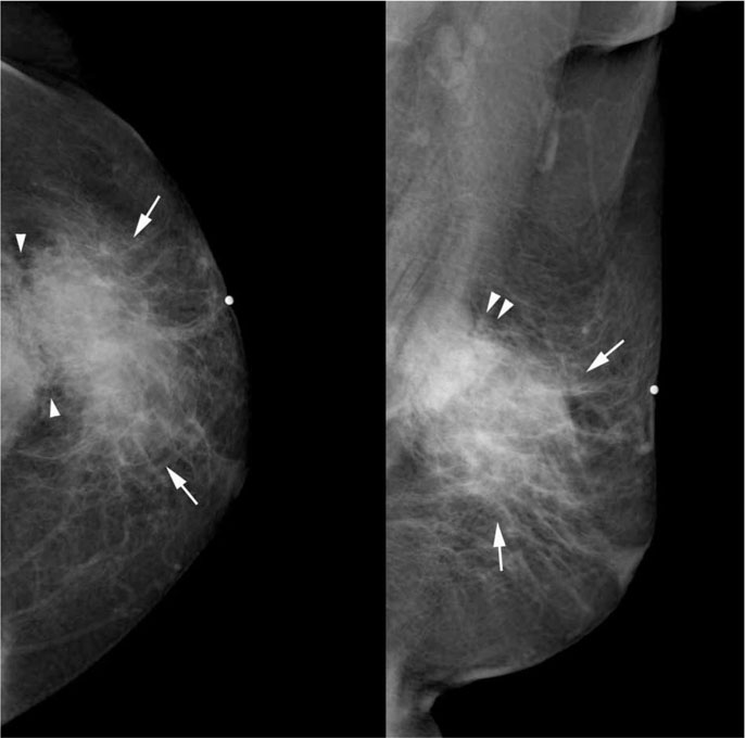
Fig. 1 Medio-lateral oblique and cranio-caudal mammographic views of the left breast shows a large, irregular shaped, hyperdense mass with an ill-defined margin and located in the upper outer quadrant, which appeared to infiltrate the pectoral muscle (arrowheads) and retract the overlying skin.
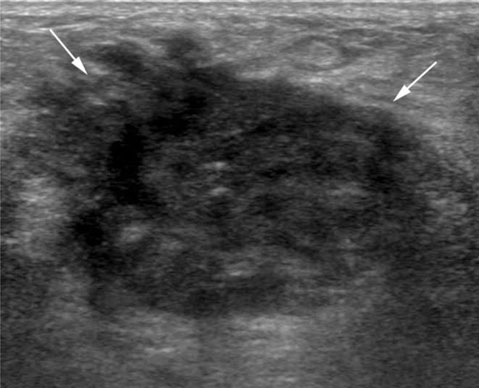
Fig. 2 Ultrasound images show an irregularly shaped hypoechoic mass with speculation.
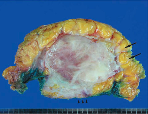
Fig. 3 Gross cross-sectional view of the pathology specimen. The pinkish-white fibrotic mass shows an infiltrating extension into the breast parenchyma (arrows) and pectoral muscle (arrowheads).

Fig. 4 Microscopic examination shows spindle cells with collagen. The spindle cells have small, pale, oval or spindly nuclei without nuclear atypia or pleomorphism, and the mitotic figures are inconspicuous (hematoxylin-eosin, ×400).
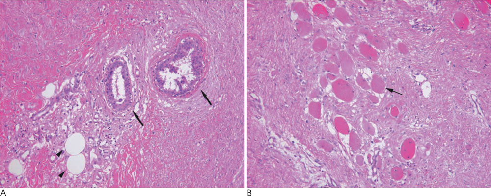
Fig. 5 Microscopic examination shows spindle cells invading and engulfing the ducts (arrows) and stromal adipose cells (arrowheads) (A, hematoxylin-eosin, ×100) and muscles (arrow) (B, hematoxylin-eosin, ×200).
Reference
-
1. Benign Mesenchymal Neoplasms. In : Rosen PP, editor. Rosen's Breast Pathology. 3rd ed. Philadelphia: Lippincott Williams & Wilkins;2009. p. 829–837.2. Rosen PP, Ernsberger D. Mammary fibromatosis. A benign spindle-cell tumor with significant risk for local recurrence. Cancer. 1989; 63:1363–1369.3. Abraham SC, Reynolds C, Lee JH, Montgomery EA, Baisden BL, Krasinskas AM, et al. Fibromatosis of the breast and mutations involving the APC/beta-catenin pathway. Hum Pathol. 2002; 33:39–46.4. Erguvan-Dogan B, Dempsey PJ, Ayyar G, Gilcrease MZ. Primary desmoid tumor (extraabdominal fibromatosis) of the breast. AJR Am J Roentgenol. 2005; 185:488–489.5. Neuman HB, Brogi E, Ebrahim A, Brennan MF, Van Zee KJ. Desmoid tumors (fibromatoses) of the breast: a 25-year experience. Ann Surg Oncol. 2008; 15:274–280.6. Nakazono T, Satoh T, Hamamoto T, Kudo S. Dynamic MRI of fibromatosis of the breast. AJR Am J Roentgenol. 2003; 181:1718–1719.7. Wargotz ES, Norris HJ, Austin RM, Enzinger FM. Fibromatosis of the breast. A clinical and pathological study of 28 cases. Am J Surg Pathol. 1987; 11:38–45.8. Ng WK, Poon CS, Lau MY, Li SM, Ma L. Actin inclusions in stromal cells of fibroepithelial tumor of breast: immunohistochemical and ultrastructural studies. Ultrastruct Pathol. 1999; 23:199–205.9. Janinis J, Patriki M, Vini L, Aravantinos G, Whelan JS. The pharmacological treatment of aggressive fibromatosis: a systematic review. Ann Oncol. 2003; 14:181–190.
- Full Text Links
-
- Actions
-
Cited
- CITED
-
- Close
- Share
-
- Similar articles
-
- Recurring Fibromatosis of Breast Following Tumorectomy: A Case Report
- Clinical Experience of Partial Resection of Desmoid Tumor and Perforated Small Bowel for Unresectable Desmoid Tumor with Small Bowel Perforation after IPAA for FAP
- Desmoid-Type Fibromatosis Associated with Silicone Breast Implants
- Desmoid Tumor of the Facet Joint: A Case Report
- CT findings of Desmoid tumor arising at Abdominai Wall
