Epithelioid Hemangioendothelioma of the Femur with Benign Cystic Appearance
- Affiliations
-
- 1Department of Radiology, Seoul National University Bundang Hospital, Seongnam, Korea. jacrad@radiol.snu.ac.kr
- 2Department of Radiology and Institute of Radiation Medicine, Seoul National University College of Medicine, Seoul, Korea.
- 3Department of Pathology, Seoul National University Bundang Hospital, Seongnam, Korea.
- 4Department of Orthopedic Surgery, Seoul National University Bundang Hospital, Seongnam, Korea.
- KMID: 2002930
- DOI: http://doi.org/10.3348/jksr.2011.65.6.607
Abstract
- An epithelioid hemangioendothelioma is an intermediate grade tumor between hemangioma and angiosarcoma that frequently shows marked enhancement because it is a vascular tumor. Herein, we describe a rare case of a malignant epithelioid hemangioendothelioma of the femur that was mistaken as a benign lesion such as a simple bone cyst or fibrous dysplasia because the tumor had a benign cystic appearance on MRI and its imaging findings showed a histopathologic correlation.
Figure
-
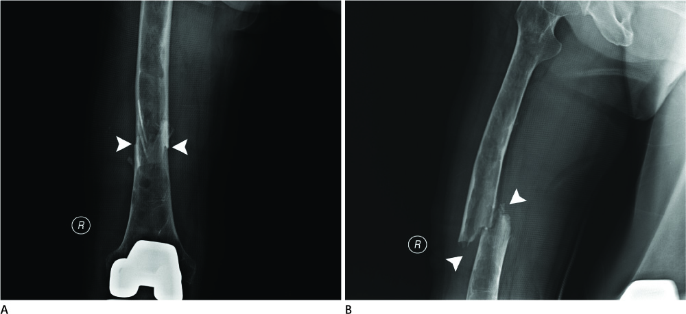
Fig. 1 On the right femur AP (A) and lateral (B) views, an elongated, radiolucent, slightly expansile lesion is seen at right femur shaft, extending from the proximal metadiaphysis to the distal metaphysic area. A pathologic fracture is noted at distal 2/3 of the femur (arrowheads). Note.-AP = anteroposterior
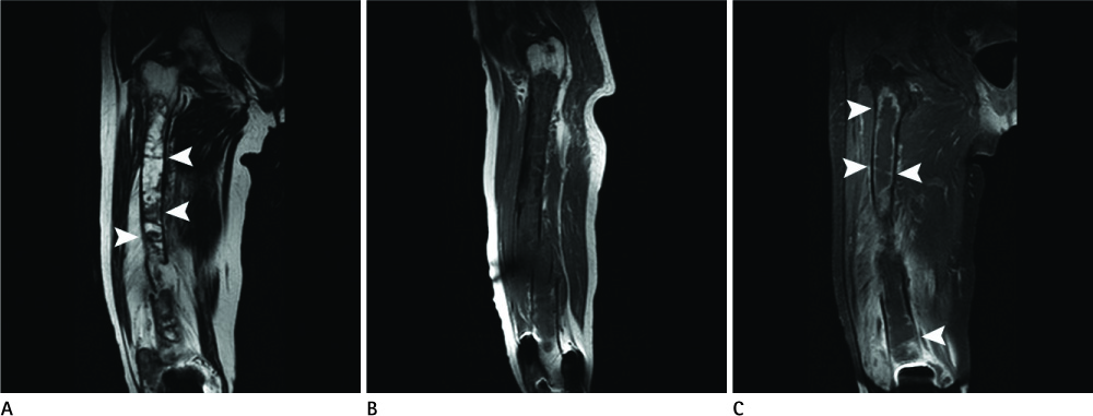
Fig. 2 MR images of right femur. A. On a coronal T2-weighted image (TR/TE = 3,734/100), the lesion extends from the proximal metadiaphysis area spanning from just below the intertrochanteric area to the distal femur up to the prosthesis. The lesion shows high signal intensity with focal intermediate to low signal intensity foci and numerous septae (arrowheads). A fracture is noted at distal two-thirds of femur. B. On a sagittal T1-weighted image (TR/TE = 421/7), the lesion is shown to have intermediate signal intensity similar to muscle. C. On a coronal post-contrast fat suppression T1-weighted image (TR/TE = 600/6), the lesion shows thin peripheral enhancement and enhancement of the septae (arrowheads). Note.-TR = repetition time, TE = echo time

Fig. 3 Histopathologic specimen images. A. On histopathologic examination, endothelial lining at the margin of the tumor and numerous vessels with RBCs (arrowheads) are seen in fragments of tissue obtained by curettage (H&E, × 10). B. Aberrant vessel formation and extravasated RBCs (arrowheads) are characteristic (H&E, × 400). C. Atypical mitoses (arrowheads) are common (H&E, × 400). Note.-RBC = red blood cell
Reference
-
1. Weiss SW, Enzinger FM. Epithelioid hemangioendothelioma: a vascular tumor often mistaken for a carcinoma. Cancer. 1982; 50:970–981.2. Vigorita VJ, Ghelman B. Vascular/Mesenchymal tumors. Orthopaedic pathology. Philadelphia: Lippincott Williams & Wilkins;1999. p. 399–400.3. Ignacio EA, Palmer KM, Mathur SC, Schwartz AM, Olan WJ. Epithelioid hemangioendothelioma of the lower extremity. Radiographics. 1999; 19:531–537.4. Larochelle O, Périgny M, Lagacé R, Dion N, Giguére C. Best cases from the AFIP: epithelioid hemangioendothelioma of bone. Radiographics. 2006; 26:265–270.5. Murphey MD, Fairbairn KJ, Parman LM, Baxter KG, Parsa MB, Smith WS. From the archives of the AFIP. Musculoskeletal angiomatous lesions: radiologic-pathologic correlation. Radiographics. 1995; 15:893–891.6. Dorfman HD, Czerniak B. Vascular lesions. Bone tumors. Mosby;1998. p. 769–795.7. Kleer CG, Unni KK, McLeod RA. Epithelioid hemangioendothelioma of bone. Am J Surg Pathol. 1996; 20:1301–1311.8. Kabukçuoğlu F, Kabukçuoğlu Y, Livaoğlu A, Ozağari A, Armağan R, Kuzgun U. [Epithelioid hemangioendothelioma of bone]. Acta Orthop Traumatol Turc. 2006; 40:324–328.
