Clinical Significance and Pathologic Outcomes of Coarse Heterogeneous Calcifications Detected on a Mammography
- Affiliations
-
- 1Department of Radiology, Korea University College of Medicine, Seoul, Korea. krcho@korea.ac.kr
- 2Department of Surgery, Korea University College of Medicine, Seoul, Korea.
- 3Department Oncology, Korea University College of Medicine, Seoul, Korea.
- KMID: 2002928
- DOI: http://doi.org/10.3348/jksr.2011.65.6.595
Abstract
- PURPOSE
To investigate the clinical significance and pathologic outcome of coarse heterogeneous calcifications (CHCs) detected on a mammography.
MATERIALS AND METHODS
A retrospective review of our institutional mammographic database revealed 65 women with CHCs. Of these, we included 27 with pathologic verification (n = 27; benign in 13, malignancy in 14). Mammograms were interpreted in terms of CHC distribution (clustered, linear, segmental, regional, or diffuse), the area of CHCs, and associated findings. We also evaluated the presence of mass, ductal change, or change of parenchymal echogenicity on ultrasound images (n = 26). We correlated and statistically analyzed the radiologic features with pathologic findings.
RESULTS
The individual distributional descriptors of CHCs predicted the risk of malignancy as follows: clustered (8/22); linear (1/2); regional (0/1); segmental (5/5). The segmental distribution predicted malignancy (p < 0.05). The CHC area in malignant lesions was larger than that of benign lesions (p < 0.05). Mammography revealed an associated mass in 2 out of 13 benign and 5 out of 14 malignancies. However, an increased risk of malignancy was not shown by the presence of an associated mass and its larger size. Ultrasound findings were not significant for predicting malignancy.
CONCLUSION
CHCs were verified as malignancy in 52% of cases, especially when characterized by segmental distribution and larger CHC areas on mammography.
Figure
-

Fig. 1 A 50-year-old woman with pathologically proven fibrocystic change. A. Mammogram shows an approximately 1 cm sized relatively oval-shaped isodense mass with clustered coarse heterogeneous calcifications (arrows) in the upper central portion of the left breast. B. On breast sonography, this lesion is about 1.2 cm in size with an indistinctly margined hypoechoic mass (arrow).

Fig. 2 A 41-year-old woman with pathologically proven ductal carcinoma in situ. A. On mammogram, linear and clustered coarse heterogeneous microcalcifications (arrows) are seen in the right mid inner portion. B. On breast ultrasonography, an indistinct oval-shaped hypoechoic mass with associated calcifications (arrows) is seen in the right breast.
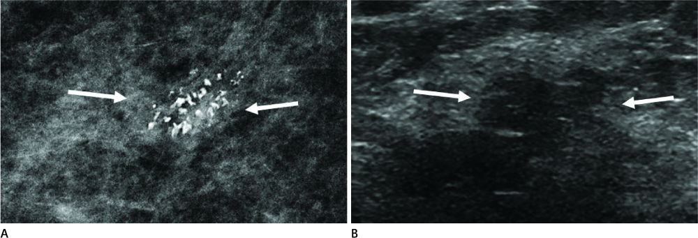
Fig. 3 A 47-year-old woman with pathologically proven microinvasive ductal carcinoma in situ. A. On magnification mammogram, coarse heterogeneous microcalcifications (arrows) are found to be clustered in right upper inner quadrant. B. On breast sonography, a 1.1 × 0.8 cm-sized hypoechoic mass (arrows) is seen at a corresponding location.
Reference
-
1. American College of Radiology. Breast imaging reporting and data system (BI-RADS). 3rd ed. Reston: American College of Radiology;1998.2. American College of Radiology. Breast imaging reporting and data system (BI-RADS). 4th ed. Reston: American College of Radiology;2003.3. Burnside ES, Ochsner JE, Fowler KJ, Fine JP, Salkowski LR, Rubin DL, et al. Use of microcalcification descriptors in BI-RADS 4th edition to stratify risk of malignancy. Radiology. 2007; 242:388–395.4. Chan KKK, Lui CY, Chu T, Chan KK, Yan AT, Wong K, et al. Stratifying risk for malignancy using microcalcification descriptors from the Breast Imaging Reporting and Data System 4th edition: experience in a single centre in Hong Kong. J HK Coll Radiol. 2009; 11:149–153.5. Tse GM, Tan PH, Pang AL, Tang AP, Cheung HS. Calcification in breast lesions: pathologists' perspective. J Clin Pathol. 2008; 61:145–151.6. Feig SA, Galkin BM, Muir HD. Evaluation of breast microcalcifications by means of optically magnified tissue specimen radiographs. Recent Results Cancer Res. 1987; 105:111–123.7. Sickles EA. Mammographic features of 300 consecutive nonpalpable breast cancers. AJR Am J Roentgenol. 1986; 146:661–663.8. Elmore JG, Armstrong K, Lehman CD, Fletcher SW. Screening for breast cancer. JAMA. 2005; 293:1245–1256.9. D'Orsi CJ. The American College of Radiology mammography lexicon: an initial attempt to standardize terminology. AJR Am J Roentgenol. 1996; 166:779–780.10. Bent CK, Bassett LW, D'Orsi CJ, Sayre JW. The positive predictive value of BI-RADS microcalcification descriptors and final assessment categories. AJR Am J Roentgenol. 2010; 194:1378–1383.11. Tanaka K, Akiyama F, Nishikawa N, Kimura K, Gomi N, Oda K, et al. Invasive carcinoma of the breast accompanied by coarse calcification. AJR Am J Roentgenol. 2009; 193:W70–W71.12. Baker JA, Kornguth PJ, Floyd CE Jr. Breast imaging reporting and data system standardized mammography lexicon: observer variability in lesion description. AJR Am J Roentgenol. 1996; 166:773–778.
- Full Text Links
-
- Actions
-
Cited
- CITED
-
- Close
- Share
-
- Similar articles
-
- Significance and diagnostic value of fine calcifications detected by mammography in female breast
- Mammography-Guided Interventional Procedure
- Imaging Findings of Metaplastic Breast Carcinoma with Chondroid Differentiation: A Case Reports
- Calcification in Thyroid Nodule and Thyroid Cancer
- Analysis of 62 Cases with Stereotaxic Breast Biopsy with a Prone Table System: Emphasis on Lesions with micro calcifications
