CT and Magnetic Resonance Imaging Findings of Psammomatoid Juvenile Ossifying Fibroma of the Middle Turbinate: A Case Report
- Affiliations
-
- 1Department of Radiology, Dongsan Medical Center, Keimyung University School of Medicine, Daegu, Korea. sklee@dsmc.or.kr
- 2Department of Pathology, Dongsan Medical Center, Keimyung University School of Medicine, Daegu, Korea.
- KMID: 2002894
- DOI: http://doi.org/10.3348/jksr.2013.68.6.449
Abstract
- Ossifying fibroma of the middle turbinate is extremely rare. We report a case of psammomatoid juvenile ossifying fibroma (PsJOF) of the middle turbinate in an 18-year-old adolescent female along with its CT, MRI and pathologic features. PsJOF of the middle turbinate may present a well-demarcated, expansile, solidly enhancing mass with focal bony destruction, which may mimic various benign and malignant neoplasms of the sinonasal area. A combination of clinical, imaging and pathologic findings is prerequisite for establishing an accurate diagnosis.
MeSH Terms
Figure
-
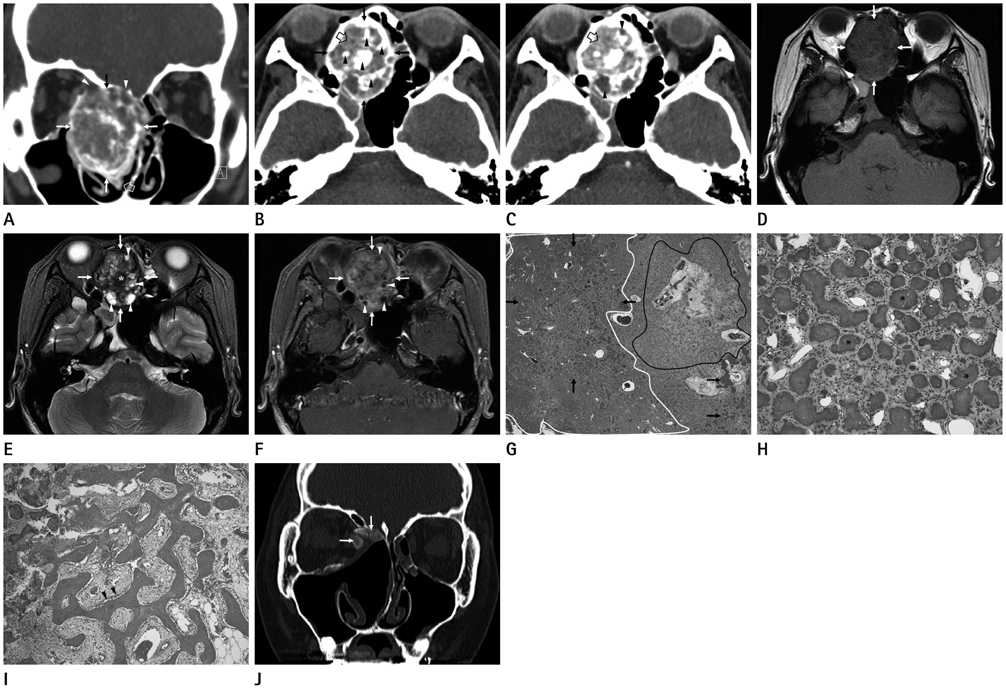
Fig. 1 CT, MRI, and pathologic features of psammomatoid juvenile ossifying fibroma of the right middle turbinate in an 18-year-old adolescent female. A. Coronal enhanced CT image shows that a well-demarcated, expansile, heterogeneous mass (arrows) originating from the right middle turbinate (open arrow) involves the right nasal cavity and ethmoid sinus. Note focal bony destruction of the right lamina papyracea and right cribriform plate (arrowheads). B. Non-enhanced CT image reveals that the tumor matrix, which consists of areas of ground-glass attenuation (open arrow), hypoattenuating areas, and irregular ossifications (arrowheads), is surrounded by a thick peripheral rim of ossification (arrows). C. Enhanced CT image demonstrates moderate enhancement of the areas of ground-glass attenuation (open arrow), while the hypoattenuating areas (arrowheads) are not enhanced. D. Axial T1-weighted image shows that the mass (arrows) is heterogeneous and hypointense compared with brain. E. Axial T2-weighted image (T2WI) with fat saturation reveals heterogeneous mass (arrows) containing multiple hyperintense cystic spaces (arrowheads). Also noted is a hypointense ossification (asterisk) in the center of the mass. F. Enhanced axial T1-weighted image demonstrates moderate enhancement of the mass (arrows). Several small non-enhancing, hypointense areas (arrowheads), which are corresponded to the hyperintense cystic spaces on fast spin echo T2WI, are seen within the moderately enhancing tumor matrix. G. Low-power photomicrograph of the specimen reveals a highly cellular area (white circle), a loose or myxoid area (black circle), and scattered psammomatoid bodies (arrows) (hematoxylin & eosin staining, × 40). H. High-power photomicrograph shows characteristic psammomatoid bodies (asterisks) and intervening fibroblastic stroma (hematoxylin & eosin staining, × 200). I. High-power photomicrograph demonstrates bony trabeculae lined by osteoblastic rimming (arrowheads). J. Bone algorithm CT image obtained 13 months after endoscopic sinus surgery reveals residual disease of ground-glass attenuation at the right medial orbital wall and cribriform plate (arrows).
Reference
-
1. Caylakli F, Buyuklu F, Cakmak O, Ozdemir H, Ozluoglu L. Ossifying fibroma of the middle turbinate: a case report. Am J Otolaryngol. 2004; 25:377–378.2. Galvan O, Gassner EM, Neher A, Gunkel AR. Fibro-osseous lesion of the middle turbinate: ossifying fibroma or fibrous dysplasia? J Laryngol Otol. 2007; 121:1201–1203.3. Pata YS, Ekici ID, Cihangiroğlu M, Doğan M, Koçak I. Ossifying fibroma of the inferior turbinate. Kulak Burun Bogaz Ihtis Derg. 2011; 21:163–166.4. Danielides V, Ingels K, Patrikakos G, de Wilde PC. Aggressive psammomatoid ossifying fibroma of the inferior turbinate and lateral nasal wall. Acta Otorhinolaryngol Belg. 2003; 57:87–90.5. Marvel JB, Marsh MA, Catlin FI. Ossifying fibroma of the mid-face and paranasal sinuses: diagnostic and therapeutic considerations. Otolaryngol Head Neck Surg. 1991; 104:803–808.6. Lund VJ. Ossifying fibroma. A case report. J Laryngol Otol. 1982; 96:1141–1147.7. Bertrand B, Eloy P, Cornelis JP, Gosseye S, Clotuche J, Gilliard C. Juvenile aggressive cemento-ossifying fibroma: case report and review of the literature. Laryngoscope. 1993; 103:1385–1390.8. London SD, Schlosser RJ, Gross CW. Endoscopic management of benign sinonasal tumors: a decade of experience. Am J Rhinol. 2002; 16:221–227.9. Post G, Kountakis SE. Endoscopic resection of large sinonasal ossifying fibroma. Am J Otolaryngol. 2005; 26:54–56.10. Han MH, Chang KH, Lee CH, Seo JW, Han MC, Kim CW. Sinonasal psammomatoid ossifying fibromas: CT and MR manifestations. AJNR Am J Neuroradiol. 1991; 12:25–30.
- Full Text Links
-
- Actions
-
Cited
- CITED
-
- Close
- Share
-
- Similar articles
-
- An expanded juvenile ossifying fibroma in maxillary sinus: a case report
- Juvenile psammomatoid ossifying fibroma of the maxilla
- A case report of juvenile active ossifying fibroma
- Juvenile Ossifying Fibroma: A Clinicopathologic Study of 8 Cases and Comparison with Craniofacial Fibro-osseous Lesions
- A Case of Psammomatoid Ossifying Fibroma of Sinonasal Tract Presenting with a Facial Deformity and Choanal Atresia
