Esthetic restoration of subgingival crown-root fractured maxillary anterior tooth using surgical extrusion
- Affiliations
-
- 1Department of Prosthodontics, Graduate School, Yonsei University, Seoul, Korea. udprostho@yuhs.ac
- 2Department of Periodontology, Graduate School, Yonsei University, Seoul, Korea.
- KMID: 2000210
- DOI: http://doi.org/10.4047/jkap.2012.50.3.204
Abstract
- Surgical extrusion, immediate extrusion following tooth luxation, is a method to preserve one's natural tooth and achieve esthetic restoration without additional periodontal surgery when subgingival dental caries or crown fracture occurs. A 16-year-old male was referred to the clinic from the department of operative dentistry for the esthetic restoration of maxillary left lateral incisor. Due to the crown to root fracture, the tooth was endodontically treated with a buccal crown length of 4 mm. When the palatal flap was elevated, the mesiopalatal cervical fracture area was situated 3 - 4 mm subgingivally. Crown lengthening was achieved through surgical extrusion. After 3 months of clinical observation and provisional restoration, the maxillary left central incisor was restored with all ceramic crown and obtained a satisfactory clinical result.
MeSH Terms
Figure
-

Fig. 1 A: Periapical radiograph at first visit. Pulp exposure is evident due to the crown-root fracture. Mature root apex can be observed, B: Periapical radiograph after root canal treatment.
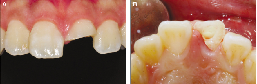
Fig. 2 Preoperative clinical examination of maxillary left central incisor reveals subgingival crown fracture. A: Labial view, B: palatal view.
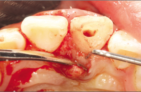
Fig. 3 Palatal view of maxillary left central incisor at palatal flap elevation. The lowest fracture line is equivalent to the alveolar bone crest.
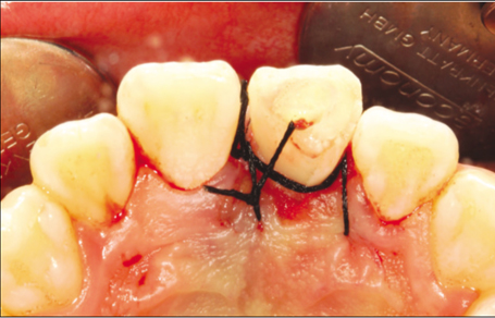
Fig. 4 The tooth is extruded approximately 3 - 4 mm and fixed only by means of suture.
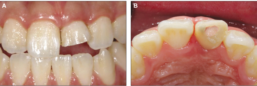
Fig. 5 1 month after surgical extrusion. A: Labial view, B: Palatal view.

Fig. 6 A: Remaining tooth structure was sufficient to obtain ferrule effect, requiring only resin core, B: Periapical radiograph 2 month postoperative.
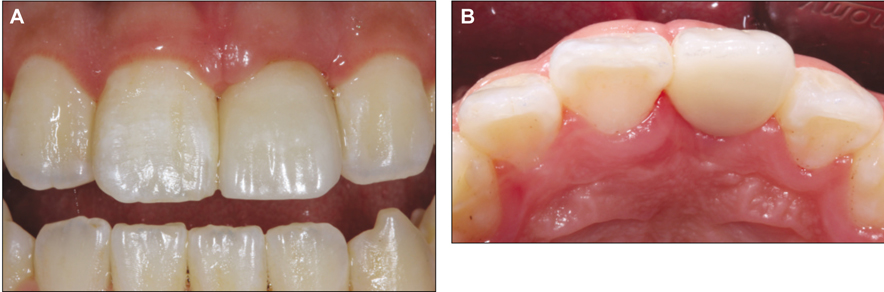
Fig. 7 1 month after delivery of lithium disilicate all ceramic crown. A: Labial view, B: Palatal view.
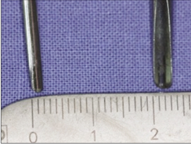
Fig. 8 Comparison photo of Periotome (left) and elevator for anterior tooth (right). Note the smaller and thinner blade of the Periotome.
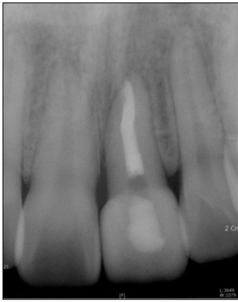
Fig. 9 Periapical radiograph 1 month after delivery of final prosthesis. The mesial emergency profile of the left central incisor is more convex than the right central incisor. Moreover the crown-to-root ratio of the left central incisor is 1:1.
Reference
-
1. Nemcovsky CE, Artzi Z, Moses O. Preprosthetic clinical crown lengthening procedures in the anterior maxilla. Pract Proced Aesthet Dent. 2001. 13:581–588.2. Kim KK. Prosthetic restoration of the maxillary anterior teeth using implantation and forced eruption: Case report. J Korean Acad Prosthodont. 2011. 49:80–86.
Article3. Bach N, Baylard JF, Voyer R. Orthodontic extrusion: periodontal considerations and applications. J Can Dent Assoc. 2004. 70:775–780.4. Kim CS, Choi SH, Chai JK, Kim CK, Cho KS. Surgical extrusion technique for clinical crown lengthening: report of three cases. Int J Periodontics Restorative Dent. 2004. 24:412–421.
Article5. Kahnberg KE. Surgical extrusion of root-fractured teeth-a follow-up study of two surgical methods. Endod Dent Traumatol. 1988. 4:85–89.
Article6. Kahnberg KE. Intra-alveolar transplantation. I. A 10-year followup of a method for surgical extrusion of root fractured teeth. Swed Dent J. 1996. 20:165–172.7. Park HK, Park JW, Suh JY, Lee JM. Surgical extrusion in aesthetic area. J Korean Acad Periodontol. 2007. 37:287–295.
Article8. Lim HC, Kim MS, Hong JY, Jung UW, Kim CS, Cho KS, Chai JK, Kim CK, Choi SH. Clinical crown lengthening procedure using surgical extrusion in esthetic region. J Korean Acad Periodontol. 2008. 38:557–564.
Article9. Diangelis AJ, Andreasen JO, Ebeleseder KA, Kenny DJ, Trope M, Sigurdsson A, Andersson L, Bourguignon C, Flores MT, Hicks ML, Lenzi AR, Malmgren B, Moule AJ, Pohl Y, Tsukiboshi M. International Association of Dental Traumatology. International Association of Dental Traumatology guidelines for the management of traumatic dental injuries: 1. Fractures and luxations of permanent teeth. Dent Traumatol. 2012. 28:2–12.
Article10. Pietrokovski J, Mersel A. The foundation for removable partial dentures. Part II. The adjacent structures. Compend Contin Educ Dent. 1982. 3:93–97.11. Kristerson L, Andreasen JO. The effect of splinting upon periodontal and pulpal healing after autotransplantation of mature and immature permanent incisors in monkeys. Int J Oral Surg. 1983. 12:239–249.
Article12. Calişkan MK, Turkun M, Gomel M. Surgical extrusion of crown-root-fractured teeth: a clinical review. Int Endod J. 1999. 32:146–151.
Article13. Koidis PT, Burch JG, Melfi RC. Clinical crown contours: contemporary view. J Am Dent Assoc. 1987. 114:792–795.
Article
- Full Text Links
-
- Actions
-
Cited
- CITED
-
- Close
- Share
-
- Similar articles
-
- Surgical extrusion of a maxillary premolar after orthodontic extrusion: a retrospective study
- A multidisciplinary approach to restore crown-root fractured maxillary central incisors: orthodontic extrusion and surgical extrusion
- Esthetic enhancement of a traumatized anterior tooth with a combination of forced eruption and tooth alignment: a case report
- Anterior esthetic improvement through orthodontic extrusive remodeling and single-unit implantation in a fractured upper lateral incisor with alveolar bone loss: A case report
- Decoronation and implant restoration of ankylosed tooth resulted from anterior avulsion: A case report
