Susceptibility Weighted MR Imaging at 3T in Patients with Occlusion of Middle Cerebral Artery : Comparison with Diffusion Weighted Imaging Score (ASPECTS)
- Affiliations
-
- 1Department of Radiology, Seoul St. Mary's Hospital, College of Medicine, The Catholic University of Korea. bumrad@catholic.ac.kr
- KMID: 2000050
- DOI: http://doi.org/10.13104/jksmrm.2011.15.3.219
Abstract
- PURPOSE
To describe the imaging findings at susceptibility weighted imaging (SWI) at 3T in patients with occlusion of middle cerebral artery, and to correlate the absence or presence of arterial bright foci in sylvian fissure, as one of their finding at SWI, with the diffusion weighted imaging (DWI) scores.
MATERIALS AND METHODS
We included 12 patients with symptomatic unilateral occlusion of middle cerebral artery. Retrospective review of SWI and DWI was done. On DWI, size of infarction was analyzed according to the ASPECTS grading system. On SWI, presence of hemorrhage, dark blooming of intravascular clot, distension of medullary or cortical vein, and absence or presence of bright arterial foci in sylvian fissure were evaluated.
RESULTS
Of 12 patients with symptomatic unilateral MCA occlusion, SWI showed dark blooming of intravascular clot in 8 patients (66.7%), distended medullary or cortical vein in 7 patients (58.3%), nonvisualization of arterial bright signal intensity in sylvian fissure in 7 patients (58.3%), and hemorrhage in one patient (8.3%). In comparison with DWI, patients with sylvian arterial bright signal intensity showed better ASPECTS score (6.4+/-4.1) than patients without arterial bright signal intensity (4.4+/-1.1), yet it was not statistically significant (p=0.267, t-test).
CONCLUSION
SWI at 3T provides added diagnostic information including site of occlusion, collateral flow by arterial bright signal intensity in sylvian fissure and early hemorrhagic transformation in patients with symptomatic MCA occlusion.
Keyword
MeSH Terms
Figure
-
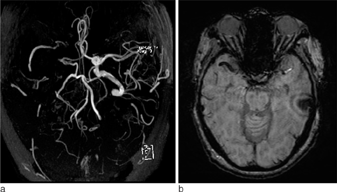
Fig. 1 Arterial blooming. 3-dimensional time-of-flight MRA (a) shows occlusion of right middle cerebral arteries. Dark signal intensity of clot in right middle cerebral artery is demonstrated on susceptibility-weighted MR image (b). The sus-ceptibility effect, attributed to clot deoxyhemoglobin content, exceeds the true MCA diameter.
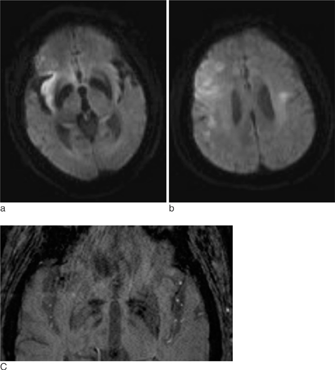
Fig. 2 Loss of arterial bright signal intensity in sylvian fissure. Diffusioin weight MR imaging (a) shows restricted diffusion area, representing acute infarction, involving right insular region. In the slice of more upper portion (b), restricted diffusion areas are also noted in the cortical and subcortical area. On magnified image of SWI (c), loss of tiny arterial bright signal foci are demonstrated in the right sylvian fissure as compared with the left side.
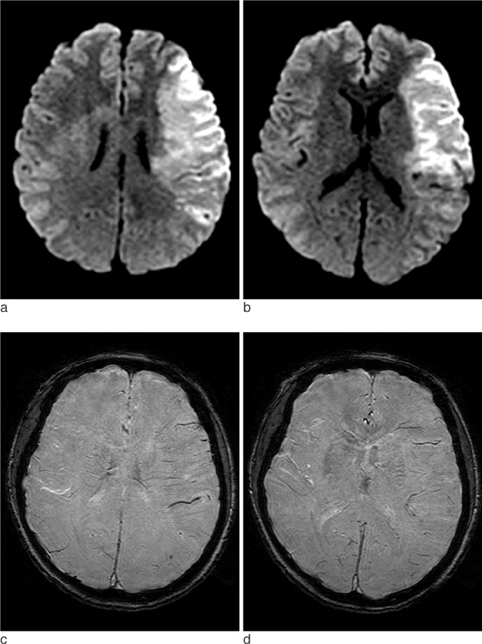
Fig. 3 Prominent veins. Diffusion weighted image (a & b) shows an acute infarct in the cortical and subcortical layer of left insula andfrontoparietal lobe. On SWI (c & d), cortical veins are prominent over the corresponding area.
Reference
-
1. Wong KS, Ng PW, Tang A, et al. Prevalence of asymptomatic intracranial atherosclerosis in high-risk patients. Neurology. 2007. 68:2035–2038.2. Adams HP Jr, Bendixen BH, Kappelle LJ, et al. Classification of subtype of acute ischemic stroke. Definitions for use in a multicenter clinical trial. TOAST. Trial of Org 10172 in Acute Stroke Treatment. Stroke. 1993. 24:35–41.3. Haacke EM, Xu Y, Cheng YC, et al. Susceptibility weighted imaging (SWI). Magn Reson Med. 2004. 52:612–618.4. Thomas B, Somasundaram S, Thamburaj K, et al. Clinical applications of susceptibility weighted MR imaging of the brain - a pictorial review. Neuroradiology. 2008. 50:105–116.5. Barber PA, Demchuk AM, Zhang J, et al. Validity and reliability of a quantitative computed tomography score in predicting outcome of hyperacute stroke before thrombolytic therapy. ASPECTS Study Group. Alberta Stroke Programme Early CT Score. Lancet. 2000. 355:1670–1674.6. Rauscher A, Sedlacik J, Barth M, et al. Magnetic susceptibility-weighted MR phase imaging of the human brain. AJNR Am J Neuroradiol. 2005. 26:736–742.7. Xu Y, Haacke EM. The role of voxel aspect ratio in determining apparent vascular phase behavior in susceptibility weighted imaging. Magn Reson Imaging. 2006. 24:155–160.8. Sehgal V, Delproposto Z, Haacke EM, et al. Clinical applications of neuroimaging with susceptibility-weighted imaging. J Magn Reson Imaging. 2005. 22:439–450.9. Flacke S, Urbach H, Keller E, et al. Middle cerebral artery (MCA) susceptibility sign at susceptibility-based perfusion MR imaging: clinical importance and comparison with hyperdense MCA sign at CT. Radiology. 2000. 215:476–482.10. Chalela JA, Haymore JB, Ezzeddine MA, et al. The hypointense MCA sign. Neurology. 2002. 58:1470.11. Hermier M, Nighoghossian N. Contribution of susceptibility-weighted imaging to acute stroke assessment. Stroke. 2004. 35:1989–1994.12. Bozzao L, Fantozzi LM, Bastianello S, et al. Early collateral blood supply and late parenchymal brain damage in patients with middle cerebral artery occlusion. Stroke. 1989. 20:735–740.13. Jung SL, Lee YJ, Ahn KJ, et al. Assessment of collateral flow with multi-phasic CT: correlation with diffusion weighted MRI in MCA occlusion. J Neuroimaging. 2011. 21:225–228.14. Yen JC, Lai YJ, Chan L. Middle cerebral artery susceptibility sign and venous prominence in acute ischemic stroke. Neurol India. 2010. 58:620–621.15. Santhosh K, Kesavadas C, Thomas B, et al. Susceptibility weighted imaging: a new tool in magnetic resonance imaging of stroke. Clin Radiol. 2009. 64:74–83.16. Moulin T, Crepin-Leblond T, Chopard JL, et al. Hemorrhagic infarcts. Eur Neurol. 1994. 34:64–77.17. Nandigam RN, Viswanathan A, Delgado P, et al. MR imaging detection of cerebral microbleeds: effect of susceptibility-weighted imaging, section thickness, and field strength. AJNR Am J Neuroradiol. 2009. 30:338–343.
- Full Text Links
-
- Actions
-
Cited
- CITED
-
- Close
- Share
-
- Similar articles
-
- Reversal of a Large Ischemic Lesion with Low Apparent Diffusion Coefficient Value by Rapid Spontaneous Recanalization
- Imaging Studies in Mouse Brain Using Clinical 3T MRI Scanner
- Hyperacute Middle Cerebral Artery Territory Infarction: Comparison of Unenhanced CT, Spin-Echo T2-weighted,Fast FLAIR, and Diffusion-weighted MR Imaging
- Initial Imaging Modality in Patients with Acute Ischemic Stroke
- Early Detection of Hyperacute Cerebral Infarction in Dogs: Comparison of Unenhanced CT, Diffusion-weighted,Spin-echo T2 - weighted, and Fast FLAIR MR Imaging
