Spontaneous Rupture of Renal Angiomyolipoma in a Female Tuberous Sclerosis Patient with Pulmonary Lymphangioleiomyomatosis
- Affiliations
-
- 1Department of Urology, College of Medicine, Korea University, Seoul, Korea. uroshim@ korea.ac.kr
- KMID: 1997135
- DOI: http://doi.org/10.4111/kju.2007.48.3.344
Abstract
- Pulmonary lymphangioleiomyomatosis (LAM) is uncommon debilitating disease, predominantly affecting women of childbearing age. Renal angiomyolipoma (AML) is the most common renal lesion in patients with tuberous sclerosis (TS), but, even without TS, is reported in up to 60% of the patients with LAM, which is frequently diagnosed several years before the LAM itself. However, it is extremely rare for renal AML and pulmonary LAM to simultaneously present in tuberous sclerosis complex. Herein, a case of spontaneous rupture of a renal AML, accompanied by pulmonary LAM, in a reproductive female with TS, managed by selective transcatheter arterial embolization (TAE), is reported.
MeSH Terms
Figure
-
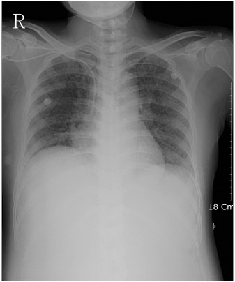
Fig. 1 Reticulonodular opacities in the parenchyma of both lungs.
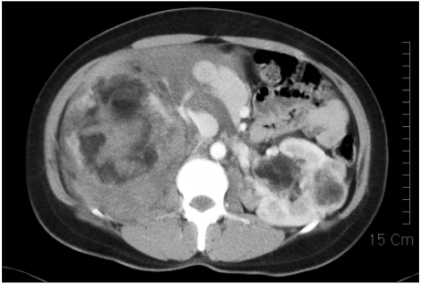
Fig. 2 Computed tomography showing a right angiomyolipoma (AML) rupture with a severe perirenal hematoma.
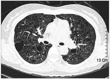
Fig. 3 Multiple cystic destruction lesions of the lung parenchyma, with a pneumothorax.
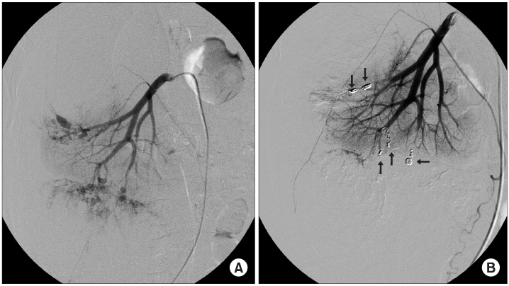
Fig. 4 (A) Right angiography showing various multiple aneurysms. (B) After transcatheter arterial embolization (TAE), the aneurysms and leakage have disappeared (arrow).
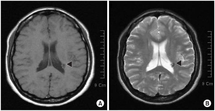
Fig. 5 (A) Multiple iso-signal nodular lesions near the lateral ventricle in the T1WI (arrowhead). (B) Multiple low-signal nodular lesions near the lateral ventricle in the T2WI (arrowhead).
Cited by 1 articles
-
Trends of Presentation and Clinical Outcome of Treated Renal Angiomyolipoma
Kyo Chul Koo, Won Tae Kim, Won Sik Ham, Jin Sun Lee, Hee Jeong Ju, Young Deuk Choi
Yonsei Med J. 2010;51(5):728-734. doi: 10.3349/ymj.2010.51.5.728.
Reference
-
1. Tuzel E, Kirkali Z, Mungan U, Culer C, Sade M. Giant angiomyolipoma associated with marked pulmonary lesions suggesting lymphangioleiomyomatosis in a patient with tuberous sclerosis. Int Urol Nephrol. 2000. 32:219–222.2. De Pauw RA, Boelaert JR, Haenebalcke CW, Matthys EG, Schurgers MS, De Vriese AS. Renal angiomyolipoma in association with pulmonary lymphangioleiomyomatosis. Am J Kidney Dis. 2003. 41:877–883.3. Tawfik O, Austenfeld M, Persons D. Multicentric renal angiomyolipoma associated with pulmonary lymphangioleiomyomatosis: case report, with histologic, immunohistochemical, and DNA content analyses. Urology. 1996. 48:476–480.4. Chang HS, Kim DI, Park JW, Kim JH, Lee DH, Park CH. A case of bilateral renal angiomyolipoma with lymph node involvement. Korean J Urol. 2001. 42:773–776.5. Cleary-Goldman J, Sanghvi AV, Nakhuda GS, Robinson JN. Conservative management of pulmonary lymphangioleiomyomatosis and tuberous sclerosis complicated by renal angiomyolipomas in pregnancy. J Matern Fetal Neonatal Med. 2004. 15:132–134.6. Carsillo T, Astrinidis A, Henske EP. Mutations in the tuberous sclerosis complex gene TSC2 are a cause of sporadic pulmonary lymphangioleiomyomatosis. Proc Natl Acad Sci USA. 2000. 97:6085–6090.7. National Heart Lung and Blood Institute. Report of Workshop on Lymphangioleiomyomatosis. AM J Respir Crit Care Med. 1999. 159:679–683.8. Dickinson M, Ruckle H, Beaghler M, Hadley HR. Renal angiomyolipoma: optimal treatment based on size and symptoms. Clin Nephrol. 1998. 49:281–286.9. Steiner MS, Goldman SM, Fishman EK, Marchall FF. The natural history of renal angiomyolipoma. J Urol. 1993. 150:1782–1786.
- Full Text Links
-
- Actions
-
Cited
- CITED
-
- Close
- Share
-
- Similar articles
-
- A Case of pulmonary lymphangioleiomyomatosis developed in tuberous sclerosis with renal angiomyolipoma
- A Case of Pulmonary Lymphangioleiomyomatosis Associated with Tuberous Sclerosis and Renal Angiomyolipoma
- A Case of Spontaneous Rupture of Renal Angiomyolipoma
- A Case of Tuberous Sclerosis with Spontaneous Rupture of Renal Angiomyolipoma
- Bilateral Renal Angiomyolipomas with Tuberous Sclerosis
