Obstet Gynecol Sci.
2015 Jan;58(1):73-76. 10.5468/ogs.2015.58.1.73.
A case of solitary fibrous tumor in the pelvis presenting massive hemorrhage during surgery
- Affiliations
-
- 1Department of Obstetrics and Gynecology, Soonchunhyang University Cheonan Hospital, Soonchunhyang University College of Medicine, Cheonan, Korea. sjeon@schmc.ac.kr
- 2Department of Obstetrics and Gynecology, Soonchunhyang University Bucheon Hospital, Soonchunhyang University College of Medicine, Bucheon, Korea.
- 3Department of Pathology, Soonchunhyang University Cheonan Hospital, Soonchunhyang University College of Medicine, Cheonan, Korea.
- KMID: 1994231
- DOI: http://doi.org/10.5468/ogs.2015.58.1.73
Abstract
- Solitary fibrous tumors (SFTs) are unique soft-tissue tumors of submesothelial origin. These tumors are mainly located in the pleural space but they can be originated within a variety of sites, including the abdomen, the pelvis, the soft tissues and the retroperitoneum. SFTs from all sites are usually benign, and the surgical resection is curative in almost all cases. According to the review of literatures, during the surgical resection, massive hemorrhage could occur due to the hypervascular nature of SFTs. This is a case report on SFT in the pelvis presenting great vessel injury, which resulted in life threatening hemorrhage during the resection of tumor. We wish this paper alerts gynecologists about the risk of massive bleeding during the resection of tumor located at adjacent to great vessels in the pelvis.
Keyword
Figure
-
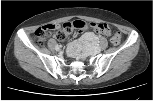
Fig. 1 (A) Contrast enhanced computed tomography; lobulated well enhancing mass in the upper-pelvic cavity is very close to left iliac vessels. (B) Same mass in the mid-pelvic cavity is located around uterus and left ovarian cyst.
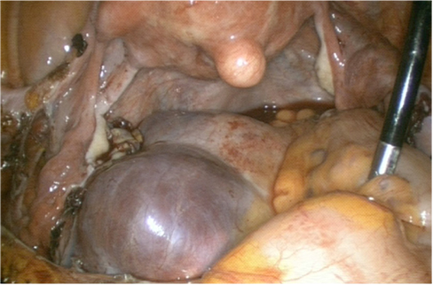
Fig. 2 Laparoscopy shows that bluish-colored huge mass is located along the sacrum in the pelvic retroperitoneum.

Fig. 3 (A) The tumor was a circumscribed mass and had many variable sized vessels (H&E, ×100). (B) The tumor was composed of high cellular spindle cells having irregular patterns and moderate cytologic atypia. Mitosis was occasionally seen (H&E, ×400). (C) The tumor cells were reactive for CD34 (×200).
Reference
-
1. Klemperer P, Coleman BR. Primary neoplasms of the pleura: a report of five cases. Am J Ind Med. 1992; 22:1–31.2. Takizawa I, Saito T, Kitamura Y, Arai K, Kawaguchi M, Takahashi K, et al. Primary solitary fibrous tumor (SFT) in the retroperitoneum. Urol Oncol. 2008; 26:254–259.3. Saint-Blancard P, Jancovici R. Solitary fibrous tumor of the retroperitoneum. Rev Med Interne. 2009; 30:181–185.4. Enzinger FM, Weiss SW. Solitary fibrous tumor. In : Enzinger FM, Weiss SW, editors. Soft tissue tumors. St. Louis: Mosby;1995. p. 1021–1031.5. England DM, Hochholzer L, McCarthy MJ. Localized benign and malignant fibrous tumors of the pleura: a clinicopathologic review of 223 cases. Am J Surg Pathol. 1989; 13:640–658.6. Hasegawa T, Matsuno Y, Shimoda T, Hasegawa F, Sano T, Hirohashi S. Extrathoracic solitary fibrous tumors: their histological variability and potentially aggressive behavior. Hum Pathol. 1999; 30:1464–1473.7. Cranshaw IM, Gikas PD, Fisher C, Thway K, Thomas JM, Hayes AJ. Clinical outcomes of extra-thoracic solitary fibrous tumours. Eur J Surg Oncol. 2009; 35:994–998.8. Wignall OJ, Moskovic EC, Thway K, Thomas JM. Solitary fibrous tumors of the soft tissues: review of the imaging and clinical features with histopathologic correlation. AJR Am J Roentgenol. 2010; 195:W55–W62.9. Wat SY, Sur M, Dhamanaskar K. Solitary fibrous tumor (SFT) of the pelvis. Clin Imaging. 2008; 32:152–156.
- Full Text Links
-
- Actions
-
Cited
- CITED
-
- Close
- Share
-
- Similar articles
-
- A Case of Solitary Fibrous Tumor in the Cheek
- Solitary Fibrous Tumor of the Adrenal Gland: A Case Report
- A Case of Intracranial Solitary Fibrous Tumor/Hemangiopericytoma Repeatedly Misdiagnosed as Hypertensive Intracerebral Hemorrhage
- Solitary Fibrous Tumor of the Renal Peripelvis
- Primary Cutaneous Solitary Fibrous Tumor on the Back
