Korean J Obstet Gynecol.
2012 Jul;55(7):454-460. 10.5468/KJOG.2012.55.7.454.
A longitudinal study of three-dimensional ultrasonographic assessment of the placenta in uncomplicated pregnancies
- Affiliations
-
- 1Department of Obstetrics and Gynecology, Kyung Hee University School of Medicine, Seoul, Korea. eui2536@hanmail.net
- KMID: 1992554
- DOI: http://doi.org/10.5468/KJOG.2012.55.7.454
Abstract
OBJECTIVE
The purpose of our study was to investigate the relationship between birth weight and 3-dimensional (3D) power Doppler histogram of the placenta in the first- and second-trimester of uncomplicated pregnancies.
METHODS
In this longitudinal study, 3D power Doppler ultrasound was performed in 54 non-smoking women with uncomplicated pregnancies at 10 to 13 weeks of gestation and subsequently at 19 to 22 weeks. A correlation and regression analysis was performed for placental volume (PV) and vascular indices versus clinical and sonographic variables.
RESULTS
All vascular indices of the second-trimester were significantly greater than values of the first-trimester. Placental vascular indices showed an incremental tendency with birth weight, which was only statistically significant when the flow index (FI) of the first-trimester was considered (P=0.02). The PV of both the first- and second-trimester has a negative correlation with FI of the second-trimester.
CONCLUSION
Our findings suggest that FI of the placenta in the first-trimester significantly affects growth of the fetus and placenta, likely reflecting the degree of placental vascularization during the first-trimester.
Keyword
Figure
-

Fig. 1 A scatter plot fitted for flow index (FI) of the first-trimester and birth weight.
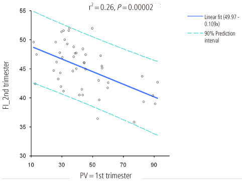
Fig. 2 A scatter plot fitted for placental volume (PV) of first-trimester and flow index (FI) of second trimester.

Fig. 3 A Bland-Altman plot of flow index intraobserver differences.
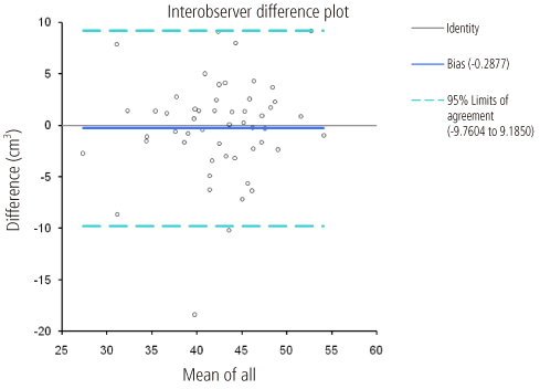
Fig. 4 A Bland-Altman plot of flow index interobserver differences.
Reference
-
1. Konje JC, Huppertz B, Bell SC, Taylor DJ, Kaufmann P. 3-dimensional colour power angiography for staging human placental development. Lancet. 2003. 362:1199–1201.2. Guimarães Filho HA, da Costa LL, Araújo Júnior E, Nardozza LM, Nowak PM, Moron AF, et al. Placenta: angiogenesis and vascular assessment through three-dimensional power Doppler ultrasonography. Arch Gynecol Obstet. 2008. 277:195–200.3. Pairleitner H, Steiner H, Hasenoehrl G, Staudach A. Three-dimensional power Doppler sonography: imaging and quantifying blood flow and vascularization. Ultrasound Obstet Gynecol. 1999. 14:139–143.4. Noguchi J, Hata K, Tanaka H, Hata T. Placental vascular sonobiopsy using three-dimensional power Doppler ultrasound in normal and growth restricted fetuses. Placenta. 2009. 30:391–397.5. Hafner E, Metzenbauer M, Stumpflen I, Waldhör T, Philipp K. First trimester placental and myometrial blood perfusion measured by 3D power Doppler in normal and unfavourable outcome pregnancies. Placenta. 2010. 31:756–763.6. Rizzo G, Capponi A, Pietrolucci ME, Capece A, Arduini D. First-trimester placental volume and vascularization measured by 3-dimensional power Doppler sonography in pregnancies with low serum pregnancy-associated plasma protein a levels. J Ultrasound Med. 2009. 28:1615–1622.7. Guiot C, Gaglioti P, Oberto M, Piccoli E, Rosato R, Todros T. Is three-dimensional power Doppler ultrasound useful in the assessment of placental perfusion in normal and growth-restricted pregnancies? Ultrasound Obstet Gynecol. 2008. 31:171–176.8. Yu CH, Chang CH, Ko HC, Chen WC, Chang FM. Assessment of placental fractional moving blood volume using quantitative three-dimensional power doppler ultrasound. Ultrasound Med Biol. 2003. 29:19–23.9. Bland JM, Altman DG. Statistical methods for assessing agreement between two methods of clinical measurement. Lancet. 1986. 1:307–310.10. de Paula CF, Ruano R, Campos JA, Zugaib M. Quantitative analysis of placental vasculature by three-dimensional power Doppler ultrasonography in normal pregnancies from 12 to 40 weeks of gestation. Placenta. 2009. 30:142–148.11. Raine-Fenning NJ, Nordin NM, Ramnarine KV, Campbell BK, Clewes JS, Perkins A, et al. Evaluation of the effect of machine settings on quantitative three-dimensional power Doppler angiography: an in-vitro flow phantom experiment. Ultrasound Obstet Gynecol. 2008. 32:551–559.12. Raine-Fenning NJ, Nordin NM, Ramnarine KV, Campbell BK, Clewes JS, Perkins A, et al. Determining the relationship between three-dimensional power Doppler data and true blood flow characteristics: an in-vitro flow phantom experiment. Ultrasound Obstet Gynecol. 2008. 32:540–550.13. Schulten-Wijman MJ, Struijk PC, Brezinka C, De Jong N, Steegers EA. Evaluation of volume vascularization index and flow index: a phantom study. Ultrasound Obstet Gynecol. 2008. 32:560–564.14. Alcázar JL. Three-dimensional power Doppler derived vascular indices: what are we measuring and how are we doing it? Ultrasound Obstet Gynecol. 2008. 32:485–487.15. Mercé LT, Barco MJ, Bau S. Reproducibility of the study of placental vascularization by three-dimensional power Doppler. J Perinat Med. 2004. 32:228–233.16. Huster KM, Haas K, Schoenborn J, McVean D, Odibo AO. Reproducibility of placental volume and vasculature indices obtained by 3-dimensional power Doppler sonography. J Ultrasound Med. 2010. 29:911–916.
- Full Text Links
-
- Actions
-
Cited
- CITED
-
- Close
- Share
-
- Similar articles
-
- The risk factors of emergency cesarean hysterectomy for placenta previa
- The association of serum placental growth factor with pregnancies complicated by preeclampsia and small for gestational age
- Preoperative Three Dimensional Ultrasonographic Evaluation of the Rotator Cuff Tear
- Altered Trophoblastic Apoptosis in Early Pregnancy Losses
- Placental apoptosis in preeclampsia
