J Korean Acad Conserv Dent.
2002 Mar;27(2):207-211. 10.5395/JKACD.2002.27.2.207.
Antimicrobial activity of eight root canal sealers before and after setting
- Affiliations
-
- 1Department of Endodontics, University of Pennsylvania, School of Dental Medicine, USA.
- 2Department of Periodontics & Microbiological Testing Laboratory, University of Pennsylvania, School of Dental Medicine, USA.
- KMID: 1987279
- DOI: http://doi.org/10.5395/JKACD.2002.27.2.207
Abstract
- No abstract available.
MeSH Terms
Figure
-
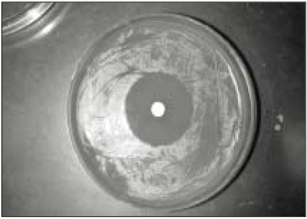
Fig. 1 Inhibition zone produced by dry AH26 against Enterococcus faecalis.
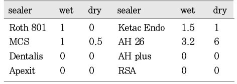
Fig. 2 Mean inhibition diameter (mm) of the wet and dry group sealers against the Enterococcus faecalis.
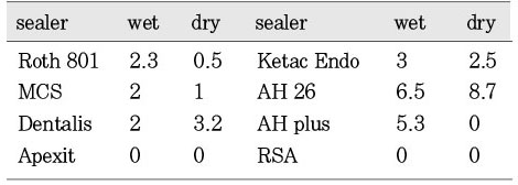
Fig. 3 Mean inhibition diameter (mm) of the wet and dry group sealers against the Staphylococcus aureus.
Reference
-
1. Kakehashi S, Stanley HR, Fizgerald RJ. The effects of surgical exposure of dental pulps in germfree and converntional rats. Oral surg. 1965. 20:340–349.
Article2. Sundqvist G. Bacteriological studies of necrotic dental pulps. Umea University Odontol Dissertation No. 7. 1976. Sweden: Umea.3. Pumarola J, Berastegui E, Brau E, Canalda C, Anta MTJ. Antimicrobial activity of seven root canal sealers. Oral Surg. 1992. 74:216–220.
Article4. Orstravik D. Antibacterial properties of root canal sealers, cements and pastes. Int Endod J. 1981. 14:125–133.
Article5. Al-Khatib ZZ, Baum RH, Morse DR, Tesilsoy C, Bhambhani S, Furst ML. The Antimicrobial effect of various endodontic sealers. Oral surg. 1990. 70:784–790.
Article6. Siqueira JF, Goncalves RB. Antibacterial activities of root canal sealers against selected anaerobic bacteria. J Endod. 1996. 22:79–80.
Article7. Kaplan AE, Picca M, Gonzalez MI, Macchi RL, Molgatini SL. Antimicrobial effect of six endodontic sealers: an in vitro evaluation. Endod Dent Traumatol. 1999. 15:42–45.
Article8. Sjögren U, Figdor D, Persson S, Sundqvist G. Influence of infection at the time of root filling on the outcome of endodontic treatment of teeth with apical periodontitis. Int Endod J. 1997. 30:297–306.
Article9. Sundqvist G, Figdor D, Persson S, Sjögren U. Microbiologic analysis of teeth with failed endodontic treatment and the outcome of conservative re-treatment. Oral Surg Oral Med Oral Pathol Oral Radiol Endod. 1998. 85:86–93.
Article10. Stevens RH, Grossman LI. Evaluation of the antimicrobial potential of calcium hydroxide as an intracanal medicament. J Endod. 1983. 9:372–374.
Article11. Spangberg LSW, Barbosa SV, Lavigne GD. AH26 releases formaldehyde. J Endod. 1993. 19:596–598.
Article12. Görduysus Ö. An evaluation of antimicrobial efficiency of endo-fill root canal sealant and filling material. J Endod. 1999. 25:652.
Article13. Duarte MAH, Demarchi AC, Giaxa MH, Kuga MC, Fraga S, Duarte de Souza LC. Evaluation of pH and calcium ion release of three root canal sealers. J Endod. 2000. 26:389–390.
Article14. Svanberg M, Mjor IA, Orstavik D. Mutans streptococci in plaque from margins of amalgam, composite, and glass-ionomer restorations. J Dent Res. 1990. 69:861–864.
Article15. Loyola-Rodriguez JP, Garcia-Godoy F, Lindquist R. Growth inhibition of glass ionomer cements on mutans streptococci. Pediatr Dent. 1994. 16:346–349.16. Ray H, Seltzer S. A new glass ionomer root canal sealer. J Endod. 1991. 17:598–603.
Article17. Friedman S, Lost C, Zarrabian M, Trope M. Evaluation of success and failure after endodontic therapy using a glass ionomer cement sealer. J Endod. 1995. 21:384–390.
Article18. Shalhav M, Fuss Z, Weiss EI. In vitro antibacterial activity of a glass ionomer endodontic sealer. J Endod. 1997. 23:616–619.
Article19. Tobias RS. Antibacterial properties of dental restorative materials: a review. Int Endod J. 1988. 21:155–160.
Article20. Cohen BI, Pagnillo MK, Musikant BL, Deutsch AS. Formaldehyde evaluation from endodontic materials. Oral Health. 1998. 88:37–39.
- Full Text Links
-
- Actions
-
Cited
- CITED
-
- Close
- Share
-
- Similar articles
-
- Calcium silicate-based root canal sealers: a literature review
- Flow characteristics and alkalinity of novel bioceramic root canal sealers
- The status of clinical trials regarding root canal sealers
- Evaluation of the radiopacity and cytotoxicity of resinous root canal sealers
- Biocompatibility and Bioactivity of Four Different Root Canal Sealers in Osteoblastic Cell Line MC3T3-El
