Left Atrial Intramural Hematoma after Removal of Atrial Myxoma: Cardiac Magnetic Resonance in the Differential Diagnosis of Intra-Cardiac Mass
- Affiliations
-
- 1Department of Internal Medicine and Cardiovascular Center, Seoul National University Hospital, Seoul, Korea. cardiman73@gmail.com
- 2Department of Radiology, Seoul National University Hospital, Seoul, Korea.
- 3Department of Thoracic and Cardiovascular Surgery, Seoul National University Hospital, Seoul, Korea.
- KMID: 1980424
- DOI: http://doi.org/10.4250/jcu.2014.22.4.205
Abstract
- Left atrial (LA) dissection is a rare entity, which is, in most cases, observed after valvular intervention. Transesophageal echocardiography (TEE) is considered to be a modality of choice in the diagnosis of LA dissection. However, LA dissection might be missed clinically in the absence of significant hemodynamic changes, and moreover physicians are occasionally reluctant to perform TEE due to its semi-invasiveness. Recently, cardiac magnetic resonance (CMR) has been introduced as a modality to perform different roles to existing imaging modalities, such as echocardiography. Given that CMR can provide information on tissue characteristics, it may give incremental information to TEE. We here present a rare case of LA dissection following LA myxoma removal, where CMR can make a correct diagnosis and guide management strategy.
MeSH Terms
Figure
-
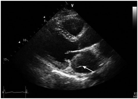
Fig. 1 Echocardiography is a diagnostic tool of choice for left atrial dissection. Left atrium occupied by a newly developed mass (arrow) was found in parasternal long axis on postoperative day 4.

Fig. 2 Postoperative thoraco-abdominal computed tomography showed a large mass having 60 Hounsfield units (arrow), which was inconclusive because of ambiguous value of Hounsfield unit.
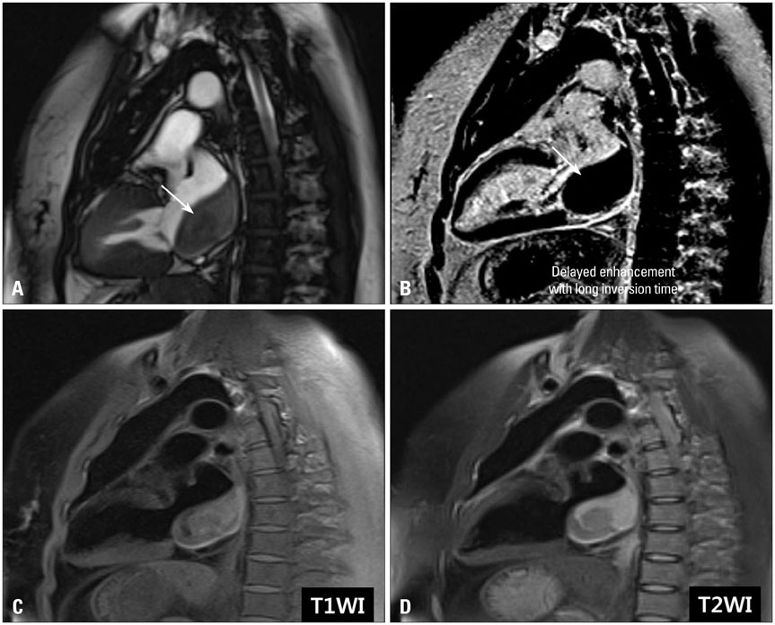
Fig. 3 Cardiac magnetic resonance performed on postoperative day 4 revealed that newly detected left atrial mass (arrow in A and B) was an intramural hematoma caused by left atrial dissection (A) showing no gadolinium enhancement (B), and heterogeneous intermediate signal intensity in T1- (C) and high T2- (D) weighted images.
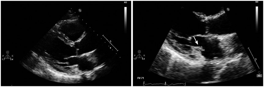
Fig. 4 Follow-up transthoracic echocardiography on postoperative day 50 confirmed that the size of left atrial mass (arrow) was getting smaller.

Fig. 5 Left atrial intramural hematoma, followed by left atrial dissection, completely disappeared on follow-up cardiac magnetic resonance imaging 5 months after index surgery. A: Sagittal view. B: Transverse view.
Reference
-
1. Martinez-Sellés M, García-Fernandez MA, Moreno M, Bermejo J, Delcán JL. Echocardiographic features of left atrial dissection. Eur J Echocardiogr. 2000; 1:147–150.
Article2. Gallego P, Oliver JM, González A, Domínguez FJ, Sanchez-Recalde A, Mesa JM. Left atrial dissection: pathogenesis, clinical course, and transesophageal echocardiographic recognition. J Am Soc Echocardiogr. 2001; 14:813–820.
Article3. Biniwale RM. Atrial septal hematoma: after minimally invasive aortic valve replacement. Tex Heart Inst J. 2010; 37:102–105.4. Osawa H, Yoshii S, Hosaka S, Suzuki S, Abraham SJ, Tada Y. Left atrial dissection after aortic valve replacement. J Thorac Cardiovasc Surg. 2003; 126:604–605.
Article5. Schecter SO, Fyfe B, Pou R, Goldman ME. Intramural left atrial hematoma complicating mitral annular calcification. Am Heart J. 1996; 132(2 Pt 1):455–457.
Article6. Cordero Lorenzana ML, López Pérez JM, Merayo Macías E, Gulías López JM, Paz Rodríguez J. [Left atrial dissection and infective endocarditis]. Rev Esp Cardiol. 1998; 51:402–403.7. Shaikh N, Rehman NU, Salazar MF, Grodman RS. Spontaneous intramural atrial hematoma presenting as a left atrial mass. J Am Soc Echocardiogr. 1999; 12:1101–1103.
Article8. Fukuhara S, Dimitrova KR, Geller CM, Hoffman DM, Ko W, Tranbaugh RF. Left atrial dissection: etiology and treatment. Ann Thorac Surg. 2013; 95:1557–1562.
Article9. Lestuzzi C, Nicolosi GL, Mimo R, Pavan D, Zanuttini D. Usefulness of transesophageal echocardiography in evaluation of paracardiac neoplastic masses. Am J Cardiol. 1992; 70:247–251.
Article10. Tasoglu I, Imren Y, Tavil Y, Zor H. Left atrial dissection following mass removal from right ventricle: non-surgical therapy. Interact Cardiovasc Thorac Surg. 2005; 4:173–174.
Article11. Delgado Jiménez JF, Rufilanchas JJ, Gómez Pajuelo C. Spontaneous left atrial haematoma. Int J Cardiol. 1991; 31:353–356.
Article12. Alvarez J, Rubio A, Mora Md, Fernández Madero G, Vivancos R, Malpartida F. [Diagnosis by magnetic resonance imaging of a case of intramural left atrial hematoma]. Rev Esp Cardiol. 2002; 55:872–874.13. Fernández-Golfín C, Jimenez Lopez-Guarch C, López Gude MJ. Left atrial wall dissection after mitral valve surgery: assessment with cardiac magnetic resonance. Magn Reson Imaging. 2011; 29:584–585.
Article14. Ortega JR, San Román JA, Rollán MJ, García A, Tejedor P, Huerta R. [Atrial hematoma in cardiac postoperative patients and the diagnostic use of transesophageal echocardiography]. Rev Esp Cardiol. 2002; 55:867–871.15. Jani S, Hecht S, Leibowitz K, Berger M. Left atrial dissection: an unusual complication of mitral valve surgery. Echocardiography. 2007; 24:443–444.
Article
- Full Text Links
-
- Actions
-
Cited
- CITED
-
- Close
- Share
-
- Similar articles
-
- A case of Right Atrial Myxoma
- Silent Left Large Atrial Myxoma: A Patient with Serial Electrocardiogram Variation
- Anesthetic Management for the Excision of Left Atrial Myxoid Sarcoma Preoperatively Diagnosed as a Left Atrial Myxoma: A case report
- Familial Cardiac Myxoma with Acromegaly(Complex Myxoma)
- Recurred Right Atrial Myxoma after Resection of Left Atrial Myxoma (Recurred Myxoma): A case report
