J Cardiovasc Ultrasound.
2011 Sep;19(3):134-139. 10.4250/jcu.2011.19.3.134.
Subclinical Myocardial Dysfunction in Metabolic Syndrome Patients without Hypertension
- Affiliations
-
- 1Department of Cardiology, Dong-A University College of Medicine, Busan, Korea. thpark65@dau.ac.kr
- 2Department of Family Medicine, Dong-A University College of Medicine, Busan, Korea.
- 3Department of Preventive Medicine, Dong-A University College of Medicine, Busan, Korea.
- KMID: 1980367
- DOI: http://doi.org/10.4250/jcu.2011.19.3.134
Abstract
- BACKGROUND
The aim of this study was to evaluate myocardial function in patients with non-hypertensive metabolic syndrome.
METHODS
We selected metabolic syndrome patients (n = 42) without evidence of hypertension and compared them to age-matched control individuals (n = 20). All patients were evaluated by two-dimensional and tissue Doppler echocardiography including tissue Doppler derived strain and strain rate measurements.
RESULTS
There were no significant differences between the two groups in mitral E and A inflow velocities or the E/A ratio. However, systolic and early diastolic myocardial velocities, and strain rate were significantly lower in patients with metabolic syndrome than in the control group (all p < 0.05). Multiple stepwise regression analyses revealed that age, waist circumference, and systolic blood pressure were independently associated with peak systolic myocardial velocity.
CONCLUSION
These results indicate that metabolic syndrome patients without hypertension may have decrease of myocardial systolic and early diastolic velocities on tissue Doppler imaging, even if they appear to have normal systolic and diastolic function on conventional echocardiography.
MeSH Terms
Figure
-
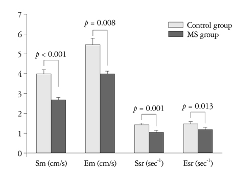
Fig. 1 Mean values of myocardial velocities and strain rate by tissue Doppler imaging in control and MS group. MS: metabolic syndrome, Sm: peak systolic, Em: early diastolic, Ssr: peak systolic, Esr: early diastolic.
Reference
-
1. Ford ES, Giles WH, Dietz WH. Prevalence of the metabolic syndrome among US adults: findings from the third National Health and Nutrition Examination Survey. JAMA. 2002. 287:356–359.
Article2. Choi KM, Kim SM, Kim YE, Choi DS, Baik SH, Lee J. International Diabetes Federation. Prevalence and cardiovascular disease risk of the metabolic syndrome using National Cholesterol Education Program and International Diabetes Federation definitions in the Korean population. Metabolism. 2007. 56:552–558.
Article3. Choi SH, Ahn CW, Cha BS, Chung YS, Lee KW, Lee HC, Huh KB, Kim DJ. The prevalence of the metabolic syndrome in Korean adults: comparison of WHO and NCEP criteria. Yonsei Med J. 2005. 46:198–205.
Article4. Isomaa B, Almgren P, Tuomi T, Forsén B, Lahti K, Nissén M, Taskinen MR, Groop L. Cardiovascular morbidity and mortality associated with the metabolic syndrome. Diabetes Care. 2001. 24:683–689.
Article5. Lakka HM, Laaksonen DE, Lakka TA, Niskanen LK, Kumpusalo E, Tuomilehto J, Salonen JT. The metabolic syndrome and total and cardiovascular disease mortality in middle-aged men. JAMA. 2002. 288:2709–2716.
Article6. Malik S, Wong ND, Franklin SS, Kamath TV, L'Italien GJ, Pio JR, Williams GR. Impact of the metabolic syndrome on mortality from coronary heart disease, cardiovascular disease, and all causes in United States adults. Circulation. 2004. 110:1245–1250.
Article7. McNeill AM, Rosamond WD, Girman CJ, Golden SH, Schmidt MI, East HE, Ballantyne CM, Heiss G. The metabolic syndrome and 11-year risk of incident cardiovascular disease in the atherosclerosis risk in communities study. Diabetes Care. 2005. 28:385–390.
Article8. Ingelsson E, Arnlöv J, Lind L, Sundström J. Metabolic syndrome and risk for heart failure in middle-aged men. Heart. 2006. 92:1409–1413.
Article9. de Simone G, Devereux RB, Chinali M, Roman MJ, Lee ET, Resnick HE, Howard BV. Metabolic syndrome and left ventricular hypertrophy in the prediction of cardiovascular events: the Strong Heart Study. Nutr Metab Cardiovasc Dis. 2009. 19:98–104.
Article10. Chinali M, Devereux RB, Howard BV, Roman MJ, Bella JN, Liu JE, Resnick HE, Lee ET, Best LG, de Simone G. Comparison of cardiac structure and function in American Indians with and without the metabolic syndrome (the Strong Heart Study). Am J Cardiol. 2004. 93:40–44.
Article11. Masugata H, Senda S, Goda F, Yoshihara Y, Yoshikawa K, Fujita N, Daikuhara H, Nakamura H, Taoka T, Kohno M. Left ventricular diastolic dysfunction as assessed by echocardiography in metabolic syndrome. Hypertens Res. 2006. 29:897–903.
Article12. de las Fuentes L, Brown AL, Mathews SJ, Waggoner AD, Soto PF, Gropler RJ, Dávila-Román VG. Metabolic syndrome is associated with abnormal left ventricular diastolic function independent of left ventricular mass. Eur Heart J. 2007. 28:553–559.
Article13. Wong CY, O'Moore-Sullivan T, Fang ZY, Haluska B, Leano R, Marwick TH. Myocardial and vascular dysfunction and exercise capacity in the metabolic syndrome. Am J Cardiol. 2005. 96:1686–1691.
Article14. Grundy SM, Cleeman JI, Daniels SR, Donato KA, Eckel RH, Franklin BA, Gordon DJ, Krauss RM, Savage PJ, Smith SC Jr, Spertus JA, Costa F. American Heart Association. National Heart, Lung, and Blood Institute. Diagnosis and management of the metabolic syndrome: an American Heart Association/National Heart, Lung, and Blood Institute Scientific Statement. Circulation. 2005. 112:2735–2752.15. Chobanian AV, Bakris GL, Black HR, Cushman WC, Green LA, Izzo JL Jr, Jones DW, Materson BJ, Oparil S, Wright JT Jr, Roccella EJ. National Heart, Lung, and Blood Institute Joint National Committee on Prevention, Detection, Evaluation, and Treatment of High Blood Pressure. National High Blood Pressure Education Program Coordinating Committee. The Seventh Report of the Joint National Committee on Prevention, Detection, Evaluation, and Treatment of High Blood Pressure: the JNC 7 report. JAMA. 2003. 289:2560–2572.
Article16. Expert Committee on the Diagnosis and Classification of Diabetes Mellitus. Report of the expert committee on the diagnosis and classification of diabetes mellitus. Diabetes Care. 2003. 26:Suppl 1. S5–S20.17. Obesity: preventing and managing the global epidemic. Report of a WHO consultation. World Health Organ Tech Rep Ser. 2000. 894:i–xii. 1–253.18. Lang RM, Bierig M, Devereux RB, Flachskampf FA, Foster E, Pellikka PA, Picard MH, Roman MJ, Seward J, Shanewise J, Solomon S, Spencer KT, St John Sutton M, Stewart W. American Society of Echocardiography's Nomenclature and Standards Committee. Task Force on Chamber Quantification. American College of Cardiology Echocardiography Committee. American Heart Association. European Association of Echocardiography, European Society of Cardiology. Recommendations for chamber quantification. Eur J Echocardiogr. 2006. 7:79–108.
Article19. Lester SJ, Tajik AJ, Nishimura RA, Oh JK, Khandheria BK, Seward JB. Unlocking the mysteries of diastolic function: deciphering the Rosetta Stone 10 years later. J Am Coll Cardiol. 2008. 51:679–689.20. Okin PM, Devereux RB, Nieminen MS, Jern S, Oikarinen L, Viitasalo M, Toivonen L, Kjeldsen SE, Julius S, Dahlöf B. Relationship of the electrocardiographic strain pattern to left ventricular structure and function in hypertensive patients: the LIFE study. Losartan Intervention For End point. J Am Coll Cardiol. 2001. 38:514–520.
Article21. Von Bibra H, Thrainsdottir IS, Hansen A, Dounis V, Malmberg K, Rydén L. Tissue Doppler imaging for the detection and quantitation of myocardial dysfunction in patients with type 2 diabetes mellitus. Diab Vasc Dis Res. 2005. 2:24–30.
Article22. Nagueh SF, Middleton KJ, Kopelen HA, Zoghbi WA, Quiñones MA. Doppler tissue imaging: a noninvasive technique for evaluation of left ventricular relaxation and estimation of filling pressures. J Am Coll Cardiol. 1997. 30:1527–1533.
Article23. Nagueh SF. Tissue Doppler imaging for the assessment of left ventricular diastolic function. J Cardiovasc Ultrasound. 2008. 16:76–79.
Article24. Park TH, Lakkis NM, Middleton KJ, Franklin J, Zoghbi WA, Quiñones MA, Spencer WH 3rd, Nagueh SF. Acute effect of nonsurgical septal reduction therapy on regional left ventricular asynchrony in patients with hypertrophic obstructive cardiomyopathy. Circulation. 2002. 106:412–415.
Article25. Takemoto Y, Pellikka PA, Wang J, Modesto KM, Cauduro S, Belohlavek M, Seward JB, Thomson HL, Khandheria B, Abraham TP. Analysis of the interaction between segmental relaxation patterns and global diastolic function by strain echocardiography. J Am Soc Echocardiogr. 2005. 18:901–906.
Article26. Palaniappan L, Carnethon MR, Wang Y, Hanley AJ, Fortmann SP, Haffner SM, Wagenknecht L. Insulin Resistance Atherosclerosis Study. Predictors of the incident metabolic syndrome in adults: the Insulin Resistance Atherosclerosis Study. Diabetes Care. 2004. 27:788–793.27. Talini E, Di Bello V, Bianchi C, Palagi C, Delle Donne MG, Penno G, Nardi C, Canale ML, Del Prato S, Mariani M, Miccoli R. Early impairment of left ventricular function in hypercholesterolemia and its reversibility after short term treatment with rosuvastatin A preliminary echocardiographic study. Atherosclerosis. 2008. 197:346–354.
Article28. Schillaci G, Pasqualini L, Verdecchia P, Vaudo G, Marchesi S, Porcellati C, de Simone G, Mannarino E. Prognostic significance of left ventricular diastolic dysfunction in essential hypertension. J Am Coll Cardiol. 2002. 39:2005–2011.
Article29. Bella JN, Palmieri V, Roman MJ, Liu JE, Welty TK, Lee ET, Fabsitz RR, Howard BV, Devereux RB. Mitral ratio of peak early to late diastolic filling velocity as a predictor of mortality in middle-aged and elderly adults: the Strong Heart Study. Circulation. 2002. 105:1928–1933.
Article
- Full Text Links
-
- Actions
-
Cited
- CITED
-
- Close
- Share
-
- Similar articles
-
- Metabolic Syndrome and Erectile Dysfunction
- Role of strain echocardiography in patients with hypertension
- Subclinical Thyroid Dysfunction in the Elderly
- The Relationship between Thyroid Function and the Risk Factors of Cardiovascular Disease at Female Medical Checkups
- Hypothyroidism and Metabolic Syndrome
