Assessment of Right Ventricular Structure and Function in Pulmonary Hypertension
- Affiliations
-
- 1Department of Cardiovascular Sciences, Imperial College of London, National Heart and Lung Institute, Hammersmith Hospital, London, UK. p.nihoyannopoulos@imperial.ac.uk
- KMID: 1980364
- DOI: http://doi.org/10.4250/jcu.2011.19.3.115
Abstract
- Right ventricular function plays an important role in determining cardiac symptoms and exercise capacity in chronic heart failure. It is known that right ventricle has complex anatomy and physiology. The purpose of this review paper is to demonstrate the best assessment of the right ventricle with current echocardiography. Echocardiography can assess sufficiently right ventricular structure and function and also suggest prognosis in pulmonary hypertension patients, especially with the use of modern imaging techniques. Finally, the new imaging modality of real time three dimensional echocardiography is interchangeable to cardiac magnetic resonance in reproducibility and accuracy.
MeSH Terms
Figure
-
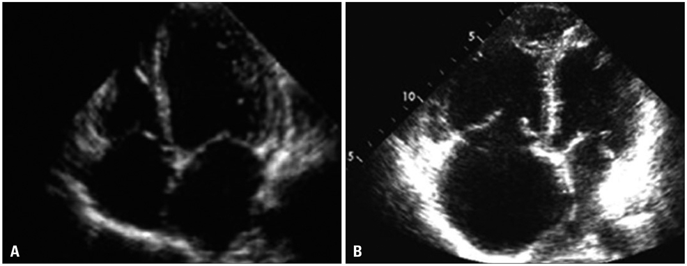
Fig. 1 A: Apical four-chamber view. Normal right-sided chambers: the right ventricle is less than one third of the size of the left ventricle. B: Apical four-chamber view. Pulmonary hypertension dilatation and hypertrophy of the right ventricle.

Fig. 2 Examples of the effects of pressure-loading (A) and volume-loading (B) of the right ventricle on the eccentricity index of the left ventricle.
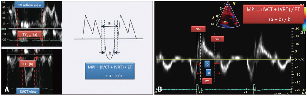
Fig. 3 Myocardial performance index. A: Tricuspid inflow from valve opening to closure (TVC-O) is the sum of isovolumic contraction time (IVCT), ejection time (ET) and isovolumic relaxation time (IVRT). The ejection time - as measured from the short axis RV outflow tract view - is subtracted from TVC-O and the result is divided by ET to give MPI. B: Measurement of MPI using tissue Doppler imaging.

Fig. 4 Calculation of right ventricle volumes and ejection fraction with 3D echocardiography.
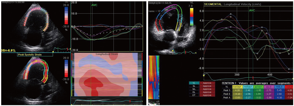
Fig. 5 Right ventricle speckle tracking.
Cited by 3 articles
-
Evaluation of Right Ventricular Systolic Function by the Analysis of Tricuspid Annular Motion in Patients with Acute Pulmonary Embolism
Jae-Hyeong Park, Jun Hyung Kim, Jae-Hwan Lee, Si Wan Choi, Jin-Ok Jeong, In-Whan Seong
J Cardiovasc Ultrasound. 2012;20(4):181-188. doi: 10.4250/jcu.2012.20.4.181.A Comparison of Different Techniques of Two-Dimensional Speckle-Tracking Strain Measurements of Right Ventricular Systolic Function in Patients with Acute Pulmonary Embolism
Jae-Hwan Lee, Jae-Hyeong Park, Kwang-In Park, Mi Joo Kim, Jun Hyung Kim, Moon Sang Ahn, Si Wan Choi, Jin-Ok Jeong, In-Whan Seong
J Cardiovasc Ultrasound. 2014;22(2):65-71. doi: 10.4250/jcu.2014.22.2.65.Assessment of Right Ventricular Function in Pulmonary Hypertension with Multimodality Imaging
Hye Sun Seo, Heon Lee
J Cardiovasc Imaging. 2018;26(4):189-200. doi: 10.4250/jcvi.2018.26.e28.
Reference
-
1. Foale R, Nihoyannopoulos P, McKenna W, Kleinebenne A, Nadazdin A, Rowland E, Smith G. Echocardiographic measurement of the normal adult right ventricle. Br Heart J. 1986. 56:33–44.
Article2. Vahanian A, Baumgartner H, Bax J, Butchart E, Dion R, Filippatos G, Flachskampf F, Hall R, Iung B, Kasprzak J, Nataf P, Tornos P, Torracca L, Wenink A. Guidelines on the management of valvular heart disease: The Task Force on the Management of Valvular Heart Disease of the European Society of Cardiology. Eur Heart J. 2007. 28:230–268.
Article3. Lang RM, Bierig M, Devereux RB, Flachskampf FA, Foster E, Pellikka PA, Picard MH, Roman MJ, Seward J, Shanewise JS, Solomon SD, Spencer KT, Sutton MS, Stewart WJ. Recommendations for chamber quantification: a report from the American Society of Echocardiography's Guidelines and Standards Committee and the Chamber Quantification Writing Group, developed in conjunction with the European Association of Echocardiography, a branch of the European Society of Cardiology. J Am Soc Echocardiogr. 2005. 18:1440–1463.
Article4. Kaul S, Tei C, Hopkins JM, Shah PM. Assessment of right ventricular function using two-dimensional echocardiography. Am Heart J. 1984. 107:526–531.
Article5. Santamore WP, Dell'Italia LJ. Ventricular interdependence: significant left ventricular contributions to right ventricular systolic function. Prog Cardiovasc Dis. 1998. 40:289–308.
Article6. López-Candales A, Dohi K, Rajagopalan N, Edelman K, Gulyasy B, Bazaz R. Defining normal variables of right ventricular size and function in pulmonary hypertension: an echocardiographic study. Postgrad Med J. 2008. 84:40–45.
Article7. Haddad F, Hunt SA, Rosenthal DN, Murphy DJ. Right ventricular function in cardiovascular disease, part I: anatomy, physiology, aging, and functional assessment of the right ventricle. Circulation. 2008. 117:1436–1448.
Article8. Haddad F, Doyle R, Murphy DJ, Hunt SA. Right ventricular function in cardiovascular disease, part II: pathophysiology, clinical importance, and management of right ventricular failure. Circulation. 2008. 117:1717–1731.
Article9. Schnittger I, Gordon EP, Fitzgerald PJ, Popp RL. Standardized intracardiac measurements of two-dimensional echocardiography. J Am Coll Cardiol. 1983. 2:934–938.
Article10. Zoghbi WA, Enriquez-Sarano M, Foster E, Grayburn PA, Kraft CD, Levine RA, Nihoyannopoulos P, Otto CM, Quinones MA, Rakowski H, Stewart WJ, Waggoner A, Weissman NJ. Recommendations for evaluation of the severity of native valvular regurgitation with two-dimensional and Doppler echocardiography. J Am Soc Echocardiogr. 2003. 16:777–802.
Article11. Fisher MR, Forfia PR, Chamera E, Housten-Harris T, Champion HC, Girgis RE, Corretti MC, Hassoun PM. Accuracy of Doppler echocardiography in the hemodynamic assessment of pulmonary hypertension. Am J Respir Crit Care Med. 2009. 179:615–621.
Article12. Kitabatake A, Inoue M, Asao M, Masuyama T, Tanouchi J, Morita T, Mishima M, Uematsu M, Shimazu T, Hori M, Abe H. Noninvasive evaluation of pulmonary hypertension by a pulsed Doppler technique. Circulation. 1983. 68:302–309.
Article13. Stein PD, Sabbah HN, Anbe DT, Marzilli M. Performance of the failing and nonfailing right ventricle of patients with pulmonary hypertension. Am J Cardiol. 1979. 44:1050–1055.
Article14. Yoshida K, Yoshikawa J, Shakudo M, Akasaka T, Jyo Y, Takao S, Shiratori K, Koizumi K, Okumachi F, Kato H. Color Doppler evaluation of valvular regurgitation in normal subjects. Circulation. 1988. 78:840–847.
Article15. Schnittger I, Gordon EP, Fitzgerald PJ, Popp RL. Standardized intracardiac measurements of two-dimensional echocardiography. J Am Coll Cardiol. 1983. 2:934–938.
Article16. Olson JM, Samad BA, Alam M. Prognostic value of pulse-wave tissue Doppler parameters in patients with systolic heart failure. Am J Cardiol. 2008. 102:722–725.
Article17. Abbas A, Lester S, Moreno FC, Srivathsan K, Fortuin D, Appleton C. Noninvasive assessment of right atrial pressure using Doppler tissue imaging. J Am Soc Echocardiogr. 2004. 17:1155–1160.
Article18. Wang Y, Gutman JM, Heilbron D, Wahr D, Schiller NB. Atrial volume in a normal adult population by two-dimensional echocardiography. Chest. 1984. 86:595–601.
Article19. Forfia PR, Fisher MR, Mathai SC, Housten-Harris T, Hemnes AR, Borlaug BA, Chamera E, Corretti MC, Champion HC, Abraham TP, Girgis RE, Hassoun PM. Tricuspid annular displacement predicts survival in pulmonary hypertension. Am J Respir Crit Care Med. 2006. 174:1034–1041.
Article20. Dujardin KS, Tei C, Yeo TC, Hodge DO, Rossi A, Seward JB. Prognostic value of a Doppler index combining systolic and diastolic performance in idiopathic-dilated cardiomyopathy. Am J Cardiol. 1998. 82:1071–1076.
Article21. Tei C, Dujardin KS, Hodge DO, Bailey KR, McGoon MD, Tajik AJ, Seward SB. Doppler echocardiographic index for assessment of global right ventricular function. J Am Soc Echocardiogr. 1996. 9:838–847.
Article22. Yeo TC, Dujardin KS, Tei C, Mahoney DW, McGoon MD, Seward JB. Value of a Doppler-derived index combining systolic and diastolic time intervals in predicting outcome in primary pulmonary hypertension. Am J Cardiol. 1998. 81:1157–1161.
Article23. Hatle L, Angelsen B. Doppler ultrasound in cardiology: physical principles and clinical applications. 1985. 2nd ed. Philadelphia: Lea & Febiger;93.24. Meluzín J, Spinarová L, Bakala J, Toman J, Krejcí J, Hude P, Kára T, Soucek M. Pulsed Doppler tissue imaging of the velocity of tricuspid annular systolic motion; a new, rapid, and non-invasive method of evaluating right ventricular systolic function. Eur Heart J. 2001. 22:340–348.
Article25. Lee CY, Chang SM, Hsiao SH, Tseng JC, Lin SK, Liu CP. Right heart function and scleroderma: insights from tricuspid annular plane systolic excursion. Echocardiography. 2007. 24:118–125.
Article26. Hammarström E, Wranne B, Pinto FJ, Puryear J, Popp RL. Tricuspid annular motion. J Am Soc Echocardiogr. 1991. 4:131–139.
Article27. Nath J, Foster E, Heidenreich PA. Impact of tricuspid regurgitation on long-term survival. J Am Coll Cardiol. 2004. 43:405–409.
Article28. Lang RM, Mor-Avi V, Sugeng L, Nieman PS, Sahn DJ. Three-dimensional echocardiography: the benefits of the additional dimension. J Am Coll Cardiol. 2006. 48:2053–2069.29. Tandri H, Daya SK, Nasir K, Bomma C, Lima JA, Calkins H, Bluemke DA. Normal reference values for the adult right ventricle by magnetic resonance imaging. Am J Cardiol. 2006. 98:1660–1664.
Article30. Mannaerts HF, van der Heide JA, Kamp O, Stoel MG, Twisk J, Visser CA. Early identification of left ventricular remodelling after myocardial infarction, assessed by transthoracic 3D echocardiography. Eur Heart J. 2004. 25:680–687.
Article31. King DL, Gopal AS, Keller AM, Sapin PM, Schröder KM. Three-dimensional echocardiography. Advances for measurement of ventricular volume and mass. Hypertension. 1994. 23:I172–I179.
Article32. van den Bosch AE, Robbers-Visser D, Krenning BJ, McGhie JS, Helbing WA, Meijboom FJ, Roos-Hesselink JW. Comparison of real-time three-dimensional echocardiography to magnetic resonance imaging for assessment of left ventricular mass. Am J Cardiol. 2006. 97:113–117.
Article33. Kjaergaard J, Petersen CL, Kjaer A, Schaadt BK, Oh JK, Hassager C. Evaluation of right ventricular volume and function by 2D and 3D echocardiography compared to MRI. Eur J Echocardiogr. 2006. 7:430–438.
Article34. Fujimoto S, Mizuno R, Nakagawa Y, Dohi K, Nakano H. Estimation of the right ventricular volume and ejection fraction by transthoracic three-dimensional echocardiography. A validation study using magnetic resonance imaging. Int J Card Imaging. 1998. 14:385–390.35. Katz J, Whang J, Boxt LM, Barst RJ. Estimation of right ventricular mass in normal subjects and in patients with primary pulmonary hypertension by nuclear magnetic resonance imaging. J Am Coll Cardiol. 1993. 21:1475–1481.
Article36. Grothues F, Moon JC, Bellenger NG, Smith GS, Klein HU, Pennell DJ. Interstudy reproducibility of right ventricular volumes, function, and mass with cardiovascular magnetic resonance. Am Heart J. 2004. 147:218–223.
Article37. Helbing WA, Rebergen SA, Maliepaard C, Hansen B, Ottenkamp J, Reiber JH, de Roos A. Quantification of right ventricular function with magnetic resonance imaging in children with normal hearts and with congenital heart disease. Am Heart J. 1995. 130:828–837.
Article38. Alfakih K, Reid S, Jones T, Sivananthan M. Assessment of ventricular function and mass by cardiac magnetic resonance imaging. Eur Radiol. 2004. 14:1813–1822.
Article39. Lorenz CH, Walker ES, Morgan VL, Klein SS, Graham TP Jr. Normal human right and left ventricular mass, systolic function, and gender differences by cine magnetic resonance imaging. J Cardiovasc Magn Reson. 1999. 1:7–21.
Article40. Pattynama PM, Lamb HJ, Van der Velde EA, Van der Geest RJ, Van der Wall EE, De Roos A. Reproducibility of MRI-derived measurements of right ventricular volumes and myocardial mass. Magn Reson Imaging. 1995. 13:53–63.
Article41. Angelini ED, Homma S, Pearson G, Holmes JW, Laine AF. Segmentation of real-time three-dimensional ultrasound for quantification of ventricular function: a clinical study on right and left ventricles. Ultrasound Med Biol. 2005. 31:1143–1158.
Article42. van Wolferen SA, Marcus JT, Boonstra A, Marques KM, Bronzwaer JG, Spreeuwenberg MD, Postmus PE, Vonk-Noordegraaf A. Prognostic value of right ventricular mass, volume, and function in idiopathic pulmonary arterial hypertension. Eur Heart J. 2007. 28:1250–1257.
Article43. Grapsa J, O'Regan DP, Pavlopoulos H, Durighel G, Dawson D, Nihoyannopoulos P. Right ventricular remodelling in pulmonary arterial hypertension with three-dimensional echocardiography: comparison with cardiac magnetic resonance imaging. Eur J Echocardiogr. 2010. 11:64–73.
Article44. Nesser HJ, Tkalec W, Patel AR, Masani ND, Niel J, Markt B, Pandian NG. Quantitation of right ventricular volumes and ejection fraction by three-dimensional echocardiography in patients: comparison with magnetic resonance imaging and radionuclide ventriculography. Echocardiography. 2006. 23:666–680.
Article45. Niemann PS, Pinho L, Balbach T, Galuschky C, Blankenhagen M, Silberbach M, Broberg C, Jerosch-Herold M, Sahn DJ. Anatomically oriented right ventricular volume measurements with dynamic three-dimensional echocardiography validated by 3-Tesla magnetic resonance imaging. J Am Coll Cardiol. 2007. 50:1668–1676.
Article46. Gopal AS, Chukwu EO, Iwuchukwu CJ, Katz AS, Toole RS, Schapiro W, Reichek N. Normal values of right ventricular size and function by real-time 3-dimensional echocardiography: comparison with cardiac magnetic resonance imaging. J Am Soc Echocardiogr. 2007. 20:445–455.
Article47. Lu X, Nadvoretskiy V, Bu L, Stolpen A, Ayres N, Pignatelli RH, Kovalchin JP, Grenier M, Klas B, Ge S. Accuracy and reproducibility of real-time three-dimensional echocardiography for assessment of right ventricular volumes and ejection fraction in children. J Am Soc Echocardiogr. 2008. 21:84–89.
Article48. Rademakers FE. 3D echocardiography: is CMR better? Eur J Echocardiogr. 2006. 7:339–340.
Article49. Badano LP, Ginghina C, Easaw J, Muraru D, Grillo MT, Lancellotti P, Pinamonti B, Coghlan G, Marra MP, Popescu BA, De Vita S. Right ventricle in pulmonary arterial hypertension: haemodynamics, structural changes, imaging, and proposal of a study protocol aimed to assess remodelling and treatment effects. Eur J Echocardiogr. 2010. 11:27–37.
Article50. Padeletti M, Cameli M, Lisi M, Zacà V, Tsioulpas C, Bernazzali S, Maccherini M, Mondillo S. Right atrial speckle tracking analysis as a novel noninvasive method for pulmonary hemodynamics assessment in patients with chronic systolic heart failure. Echocardiography. 2011. 28:658–664.
Article51. Abbas AE, Fortuin FD, Schiller NB, Appleton CP, Moreno CA, Lester SJ. A simple method for noninvasive estimation of pulmonary vascular resistance. J Am Coll Cardiol. 2003. 41:1021–1027.
Article52. British Cardiac Society Guidelines and Medical Practice Committee, and approved by the British Thoracic Society and the British Society of Rheumatology. Recommendations on the management of pulmonary hypertension in clinical practice. Heart. 2001. 86:Suppl 1. I1–I13.53. Raymond RJ, Hinderliter AL, Willis PW, Ralph D, Caldwell EJ, Williams W, Ettinger NA, Hill NS, Summer WR, de Boisblanc B, Schwartz T, Koch G, Clayton LM, Jöbsis MM, Crow JW, Long W. Echocardiographic predictors of adverse outcomes in primary pulmonary hypertension. J Am Coll Cardiol. 2002. 39:1214–1219.
Article54. Ghio S, Klersy C, Magrini G, D'Armini AM, Scelsi L, Raineri C, Pasotti M, Serio A, Campana C, Viganò M. Prognostic relevance of the echocardiographic assessment of right ventricular function in patients with idiopathic pulmonary arterial hypertension. Int J Cardiol. 2010. 140:272–278.
Article
- Full Text Links
-
- Actions
-
Cited
- CITED
-
- Close
- Share
-
- Similar articles
-
- Assessing Right Ventricular Function: The Role of Echocardiography in a Murine Model of Pulmonary Hypertension
- Acute Myocardial Infarction in 14-Year-Old Male of Primary Pulmonary Hypertension with Left Ventricular Hypertrophy : A Case Report
- Assessment of Right Ventricular Function in Pulmonary Hypertension with Multimodality Imaging
- Echocardiographic Evaluation of Right Ventricular Diastolic Function in Patients with Chronic Obstructive Pulmonary Disease
- Anesthetic Management in a Patient with Severe Primary Pulmonary Hypertension with Right Ventricular Dysfunction: A case report
