Unusual Presentation Chronic Pulmonary Embolism due to Calcified Right Ventricular Mass
- Affiliations
-
- 1Department of Cardiology, Cardiovascular Center, Myongji Hospital, Kwandong University College of Medicine, Goyang, Korea. princette@paran.com
- KMID: 1980358
- DOI: http://doi.org/10.4250/jcu.2011.19.2.91
Abstract
- Cardiac calcified amorphous tumors (CATs) can arise in all four chambers of the heart. Cardiac CATs can cause diverse symptoms according to their locations, and mass or embolic effects. Pulmonary emboli arising from cardiac CATs have been reported, but the true incidence is unknown due to their rarity. Herein we report a rare case with diffuse CATs in the right ventricle which caused a calcific pulmonary embolism and right-sided heart failure. Echocardiography, chest non-contrast computed tomography, and cardiac magnetic resonance imaging helped us diagnose the CATs. We recommend the usefulness of a multimodality imaging approach to characterize intracardiac masses and their complications accurately.
MeSH Terms
Figure
-
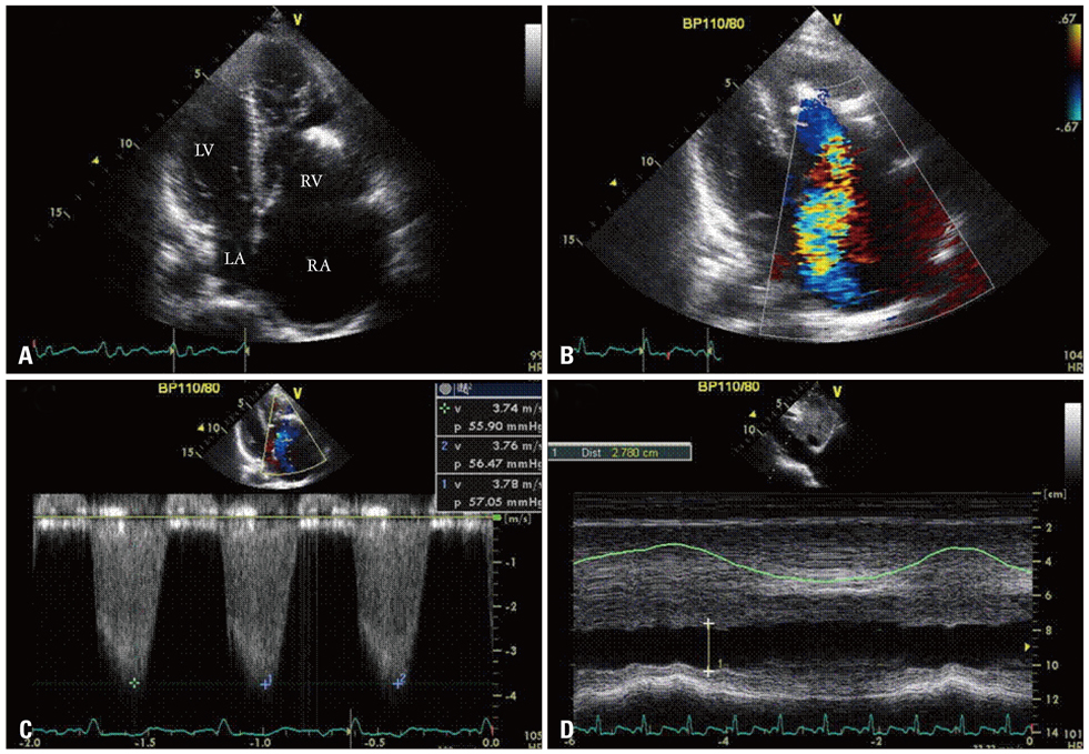
Fig. 1 Transthoracic echocardiogram. Diffuse calcified mass affecting the tricuspid chordal apparatus and the free wall of the right ventricle (A), which caused significant tricuspid regurgitation (B). The systolic pulmonary artery pressure was about 65 mmHg considering a tricuspid jet velocity of 3.76 m/s (C) and dilated inferior vena cava of 2.78 cm (D). RA: right atrium, RV: right ventricle, LV: left ventricle, LA: left atrium.
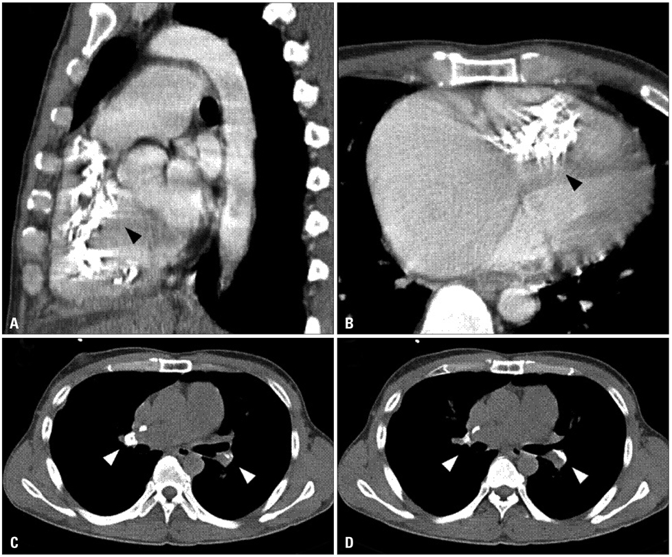
Fig. 2 Chest computed tomogram non-contrast imaging. The black arrowheads indicate calcified right ventricular amorphous tumor (A and B), and the white arrowheads indicate multiple pulmonary calcified emboli obstructing multiple pulmonary segmental arteries bilaterally (C and D).
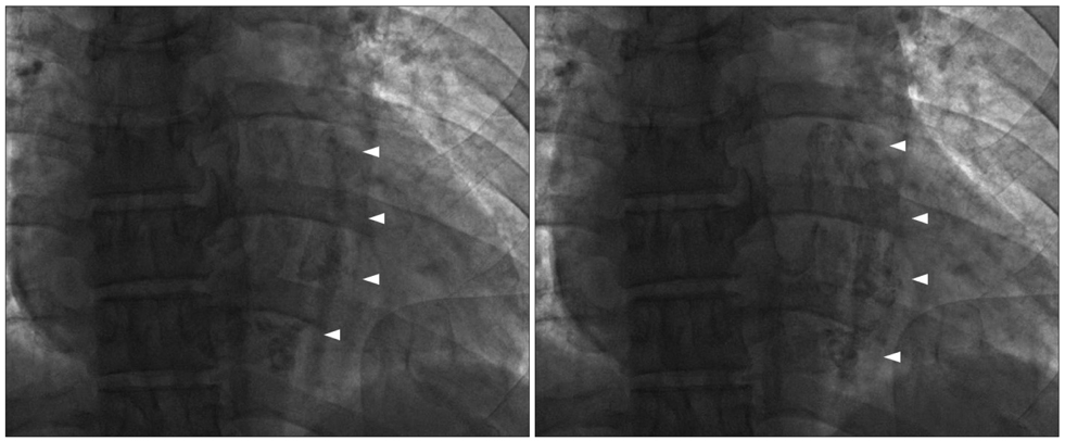
Fig. 3 Cardiac fluoroscopic imaging. The white arrowheads indicate an irregular-shaped calcified mass in the right ventricle which was changed its shape and size during to the cardiac cycle.

Fig. 4 Cardiac magnetic resonance imaging. The white arrows indicate the tubular linear calcified mass extending from just below the tricuspid valve annulus to the right ventricular outflow tract.
Reference
-
1. Ho HH, Min JK, Lin F, Wong SC, Bergman G. Images in cardiovascular medicine. Calcified amorphous tumor of the heart. Circulation. 2008. 117:e171–e172.2. Chaowalit N, Dearani JA, Edwards WD, Pellikka PA. Calcified right ventricular mass and pulmonary embolism in a previously healthy young woman. J Am Soc Echocardiogr. 2005. 18:275–277.
Article3. Tsuchihashi K, Nozawa A, Marusaki S, Moniwa N, Oh-numa Y, Kuno A, Takagi S, Takizawa H, Ura N, Shimamoto K. Mobile intracardiac calcinosis: a new risk of thromboembolism in patients with haemodialysed end stage renal disease. Heart. 1999. 82:638–640.
Article4. Reynolds C, Tazelaar HD, Edwards WD. Calcified amorphous tumor of the heart (cardiac CAT). Hum Pathol. 1997. 28:601–606.
Article5. Fealey ME, Edwards WD, Reynolds CA, Pellikka PA, Dearani JA. Recurrent cardiac calcific amorphous tumor: the CAT had a kitten. Cardiovasc Pathol. 2007. 16:115–118.
Article6. Morishima A, Sasahashi N, Ueyama K. [Calcified amorphous tumors with excision in hemodialysis patients: report of 2 cases]. Kyobu Geka. 2006. 59:851–854.7. Gutiérrez-Barrios A, Muriel-Cueto P, Lancho-Novillo C, Sancho-Jaldón M. Calcified amorphous tumor of the heart. Rev Esp Cardiol. 2008. 61:892–893.
Article8. Habib A, Friedman PA, Cooper LT, Suleiman M, Asirvatham SJ. Cardiac calcified amorphous tumor in a patient presenting for ventricular tachycardia ablation: intracardiac echocardiogram diagnosis and management. J Interv Card Electrophysiol. 2010. 29:175–178.
Article9. Iqbal MB, Stavri G, Mittal T, Khaghani A. A calcified cardiac mass. Int J Cardiol. 2007. 115:e126–e128.
Article10. Nowrangi SK, Ammash NM, Edwards WD, Breen JF, Edmonson JH. Calcified left ventricular mass: unusual clinical, echocardiographic, and computed tomographic findings of primary cardiac osteosarcoma. Mayo Clin Proc. 2000. 75:743–747.
Article
- Full Text Links
-
- Actions
-
Cited
- CITED
-
- Close
- Share
-
- Similar articles
-
- A Case of Pulmonary Embolism Due to Metastatic Chondrosarcoma
- A Case of Consecutive Right and Left Ventricular Dysfunction
- A Case of Massive Pulmonary Embolism Masked by a Ventricular Septal Defect
- Wilms' Tumor Presenting as Sudden Death due to Pulmonary Tumor Embolism
- Left Ventricular Myxoma Associated Acute Pulmonary Embolism
