Korean J Pain.
2013 Apr;26(2):203-206. 10.3344/kjp.2013.26.2.203.
Inadvertent Dural Puncture during Caudal Approach by the Introducer Needle for Epidural Adhesiolysis Caused by Anatomical Variation
- Affiliations
-
- 1Department of Anesthesiology and Pain Medicine, Ajou University College of Medicine, Suwon, Korea. yjyhy@hanmail.net
- KMID: 1978671
- DOI: http://doi.org/10.3344/kjp.2013.26.2.203
Abstract
- There have been reports of abnormalities in the lumbosacral region involving a lower-than-normal termination of the dural sac, which is caused by disease or anatomical variation. Inadvertent dural puncture or other unexpected complications can occur during caudal epidural block or adhesiolysis in patients with these variations, but only a small number of case reports have described this issue. We report a case of dural puncture by the introducer needle before attempting caudal epidural adhesiolysis, which occurred even though the needle was not advanced upward after penetrating the sacrococcygeal ligament. Dural puncture was caused by a morphological abnormality in the lumbosacral region, with no pathological condition; the dural sac terminal was located more distally than normal. However, dural puncture could have been prevented if we had checked for such an abnormality in the magnetic resonance imaging (MRI) taken before the procedure.
Keyword
Figure
-
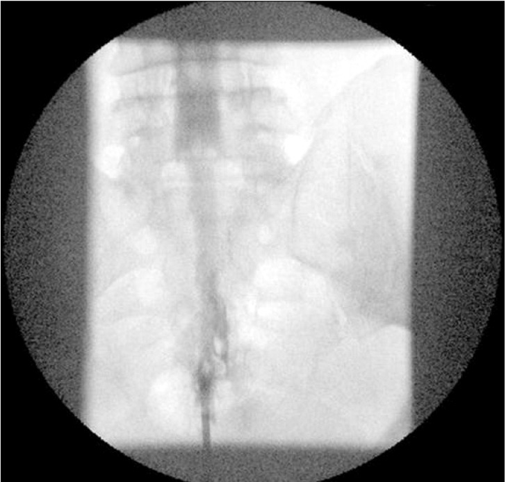
Fig. 1 Contrast dye injection to the introducer needle showed a myelogram in the anteroposterior view.

Fig. 2 Sagittal T1-weighted (A) and T2-weighted (B) MR images show extension of dural sac to the lower level of sacrum.
Cited by 1 articles
-
A Comparison of Two Techniques for Ultrasound-guided Caudal Injection: The Influence of the Depth of the Inserted Needle on Caudal Block
A Ram Doo, Jin Wan Kim, Ji Hye Lee, Young Jin Han, Ji Seon Son
Korean J Pain. 2015;28(2):122-128. doi: 10.3344/kjp.2015.28.2.122.
Reference
-
1. Meyer RJ. Dural puncture via the sacral hiatus. Anaesthesia. 1984; 39:610. PMID: 6742403.
Article2. Park BC, Kim BS, Hwang WJ, Lee J, Moon DE. Anatomical variations which can result in inadvertent dural puncture when performing caudal block: a report of 3 cases. Korean J Anesthesiol. 2006; 50:332–336.
Article3. Kang MS, Lim YJ, Lee SC. Sacral meningeal cyst detected during caudal epidural block. J Korean Pain Soc. 1999; 12:258–262.4. Joo J, Kim J, Lee J. The prevalence of anatomical variations that can cause inadvertent dural puncture when performing caudal block in Koreans: a study using magnetic resonance imaging. Anaesthesia. 2010; 65:23–26. PMID: 19922508.
Article5. Lubenow T, Keh-Wong E, Kristof K, Ivankovich O, Ivankovich AD. Inadvertent subdural injection: a complication of an epidural block. Anesth Analg. 1988; 67:175–179. PMID: 3341567.6. Racz GB, Holubec JT. Lysis of adhesion in the epidural space. Techniques of neurolysis. 1989. Boston: Kluwer Academic;p. 57–72.7. Evison G, Windsor P, Duck F. Myelographic features of the normal sacral sac. Br J Radiol. 1979; 52:777–780. PMID: 509023.
Article8. Larsen JL, Olsen KO. Radiographic anatomy of the distal dural sac. A myelographic investigation of dimensions and termination. Acta Radiol. 1991; 32:214–219. PMID: 1829618.9. Macdonald A, Chatrath P, Spector T, Ellis H. Level of termination of the spinal cord and the dural sac: a magnetic resonance study. Clin Anat. 1999; 12:149–152. PMID: 10340453.
Article10. Aggarwal A, Kaur H, Batra YK, Aggarwal AK, Rajeev S, Sahni D. Anatomic consideration of caudal epidural space: a cadaver study. Clin Anat. 2009; 22:730–737. PMID: 19637298.
Article11. Waldman JD. Pain management. 2011. 2nd ed. Philadelphia: Elsevier Saunders;p. 248–256.
- Full Text Links
-
- Actions
-
Cited
- CITED
-
- Close
- Share
-
- Similar articles
-
- Dural Puncture by Introducer when Performing Spinal Anesthesia -A case report-
- Anatomical Variations Which Can Result in Inadvertent Dural Puncture When Performing Caudal Block: A report of 3 cases
- The Effects of Needle Type, Gauge, Insertion Depth and the Use of Introducer Needle on Spinal or Epidural Needle Deflection
- Inadvertent Dural Puncture during Epidural Block
- Inadvertent Dural Puncture during Epidural Block
