Korean J Orthod.
2012 Dec;42(6):329-334. 10.4041/kjod.2012.42.6.329.
Prevalence of incidental maxillary sinus findings in Italian orthodontic patients: a retrospective cone-beam computed tomography study
- Affiliations
-
- 1Department of Orthodontics, School of Dentistry, University of Padua, Padua, Italy.
- 2Department of Orthodontics, School of Dentistry, University of Bologna, Bologna, Italy. serena.incerti2@unibo.it
- 3Department of Orthodontics, School of Dentistry, University of Ferrara, Ferrara, Italy.
- 4Department of Pedodontics, School of Dentistry, University of Padua, Padua, Italy.
- KMID: 1976685
- DOI: http://doi.org/10.4041/kjod.2012.42.6.329
Abstract
OBJECTIVES
To determine the prevalence of incidental maxillary sinus findings in a large sample of orthodontic patients by cone-beam computed tomography (CBCT) with a wide field of view and assess the relationships of such abnormalities with age and gender.
METHODS
Five hundred thirteen CBCT scans obtained for orthodontic diagnosis and treatment planning in a Northern Italian population (N = 513; 292 female and 221 male subjects; 1,026 maxillary sinuses) were studied. The frequencies of pseudocysts and mucosal thickening of the maxillary sinus were recorded. Logistic regression analysis was used to determine the influence of age and gender on these abnormalities.
RESULTS
Pseudocysts were detected in 52 patients (10.1%) and 59 sinuses (5.75%). Mucosal thickening was observed in 206 patients (40.1%) and 258 sinuses (25.1%). Gender and age were significantly associated with pseudocysts (p = 0.027) and mucosal thickening (p < 0.001), respectively.
CONCLUSIONS
Half of the orthodontic patients had incidental maxillary sinus findings. Men were more likely to show pseudocysts, and older patients (aged 41 - 60 years) were more likely to show mucosal thickening.
MeSH Terms
Figure
-
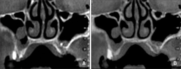
Figure 1 CBCT scans with incidental findings of pseudocysts associated with mucosal thickening in the right maxillary sinus.
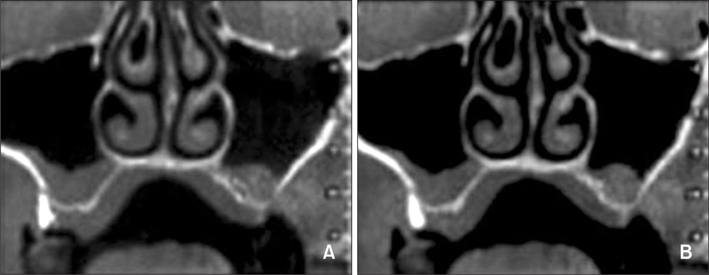
Figure 2 CBCT scans with incidental findings of mucosal thickening in the right maxillary sinus.
Reference
-
1. Horner K, Islam M, Flygare L, Tsiklakis K, Whaites E. Basic principles for use of dental cone beam computed tomography: consensus guidelines of the European Academy of Dental and Maxillofacial Radiology. Dentomaxillofac Radiol. 2009. 38:187–195.
Article2. Cha JY, Mah J, Sinclair P. Incidental findings in the maxillofacial area with 3-dimensional cone-beam imaging. Am J Orthod Dentofacial Orthop. 2007. 132:7–14.
Article3. Rogers SA, Drage N, Durning P. Incidental findings arising with cone beam computed tomography imaging of the orthodontic patient. Angle Orthod. 2011. 81:350–355.
Article4. Ritter L, Lutz J, Neugebauer J, Scheer M, Dreiseidler T, Zinser MJ, et al. Prevalence of pathologic findings in the maxillary sinus in cone-beam computerized tomography. Oral Surg Oral Med Oral Pathol Oral Radiol Endod. 2011. 111:634–640.
Article5. Pazera P, Bornstein MM, Pazera A, Sendi P, Katsaros C. Incidental maxillary sinus findings in orthodontic patients: a radiographic analysis using cone-beam computed tomography (CBCT). Orthod Craniofac Res. 2011. 14:17–24.
Article6. Carter LC, Calamel A, Haller A, Aguirre A. Seasonal variation in maxillary antral pseudocysts in a general clinic population. Dentomaxillofac Radiol. 1998. 27:22–24.
Article7. Carter L, Farman AG, Geist J, Scarfe WC, Angelopoulos C, Nair MK, et al. American Academy of Oral and Maxillofacial Radiology. American Academy of Oral and Maxillofacial Radiology executive opinion statement on performing and interpreting diagnostic cone beam computed tomography. Oral Surg Oral Med Oral Pathol Oral Radiol Endod. 2008. 106:561–562.
Article8. Phothikhun S, Suphanantachat S, Chuenchompoonut V, Nisapakultorn K. Cone-beam computed tomographic evidence of the association between periodontal bone loss and mucosal thickening of the maxillary sinus. J Periodontol. 2012. 83:557–564.
Article9. Vallo J, Suominen-Taipale L, Huumonen S, Soikkonen K, Norblad A. Prevalence of mucosal abnormalities of the maxillary sinus and their relationship to dental disease in panoramic radiography: results from the Health 2000 Health Examination Survey. Oral Surg Oral Med Oral Pathol Oral Radiol Endod. 2010. 109:e80–e87.
Article10. Bondemark L, Jeppsson M, Lindh-Ingildsen L, Rangne K. Incidental findings of pathology and abnormality in pretreatment orthodontic panoramic radiographs. Angle Orthod. 2006. 76:98–102.11. Gordts F, Clement PA, Buisseret T. Prevalence of sinusitis signs in a non-ENT population. ORL J Otorhinolaryngol Relat Spec. 1996. 58:315–319.
Article12. Havas TE, Motbey JA, Gullane PJ. Prevalence of incidental abnormalities on computed tomographic scans of the paranasal sinuses. Arch Otolaryngol Head Neck Surg. 1988. 114:856–859.
Article13. Bósio JA, Tanaka O, Rovigatti E, de Gruner SK. The incidence of maxillary sinus retention cysts in orthodontic patients. World J Orthod. 2009. 10:e7–e8.14. Kravitz ND, Kusnoto B. Risks and complications of orthodontic miniscrews. Am J Orthod Dentofacial Orthop. 2007. 131:4 Suppl. S43–S51.
Article15. Papadopoulos MA, Papageorgiou SN, Zogakis IP. Clinical effectiveness of orthodontic miniscrew implants: a meta-analysis. J Dent Res. 2011. 90:969–976.
Article16. Gracco A, Tracey S, Baciliero U. Miniscrew insertion and the maxillary sinus: an endoscopic evaluation. J Clin Orthod. 2010. 44:439–443.17. Graham JW, Cope JB. Cope JB, editor. Potential complications with OrthoTADs: classification, prevention, and treatment. OrthoTADs: The clinical guide and atlas. 2007. Dallas: Under Dog Media LP;119–134.
- Full Text Links
-
- Actions
-
Cited
- CITED
-
- Close
- Share
-
- Similar articles
-
- Prevalence of incidental paranasal sinus opacification in dental paediatric patients
- A case report of incidental finding of fungus ball on CBCT of maxillary sinus in treatment planning of dental implant
- Prevalence of incidental paranasal sinus opacification in an adult dental population
- Comparison of panoramic radiography and cone beam computed tomography for assessing the relationship between the maxillary sinus floor and maxillary molars
- Incidental finding of metastatic malignancy involving the sphenoid sinus on a cone-beam computed tomographic scan: A case report
