Histologic assessment of the biological effects after speedy surgical orthodontics in a beagle animal model: a preliminary study
- Affiliations
-
- 1Department of Dentistry, School of Medicine, Ajou University, Korea.
- 2Department of Craniofacial Orthodontics, Childrens' Hospital of Phildelphia, USA.
- 3Department of Orthodontics, School of Dentistry, Kyung Hee University, Korea. bravortho@hanmail.net
- KMID: 1975393
- DOI: http://doi.org/10.4041/kjod.2011.41.5.361
Abstract
OBJECTIVE
Speedy surgical orthodontics (SSO), an innovative orthodontic treatment, involves the application of orthopedic forces against temporary skeletal anchorage devices following perisegmental corticotomy to induce movement of specific dental segments. Herein, we report the biological effects of SSO on the teeth and periodontal structures.
METHODS
Five beagle dogs were divided into 2 groups and their 6 maxillary incisors were retracted en masse by applying 500 g orthopedic force against a single palatal mini-plate. Retraction was performed without and with perisegmental corticotomy in groups I and II, respectively. All animals were killed on the 70th day, and their periodontal structures were processed for histologic analyses and scanning electronic microscopy (SEM). The linear distance between the third maxillary incisor and canine was used as a benchmark to quantify the retraction amount.
RESULTS
Retraction was markedly faster and retraction amount greater in group II than in Group I. Surprisingly, Group II did not show any root resorption despite extensive retraction, while Group I showed prominent root surface irregularities. Similarly, SEM showed multiple resorption lacunae in Group I, but not in Group II.
CONCLUSIONS
SSO is an effective and favorable orthodontic approach for major en masse retraction of the maxillary anterior teeth.
Keyword
MeSH Terms
Figure
-

Fig. 1 Surgical cuts for perisegmental corticotomy between the maxillary 3rd incisor and canine. A, Palatal corticotomy; B, labial corticotomy.
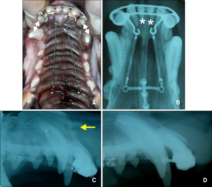
Fig. 2 A, Intraoral photograph and; B, occlusal radiograph of the orthodontic appliances used for tooth movement immediately after force application using the modified C-lingual retractor and C-plate combined appliance; C, lateral cephalogram at the time of initial force application; D, lateral cephalogram after retraction. Distance measured (white arrow) and teeth used for histologic evaluation (*) are shown, and the arrow indicates the corticotomy site.
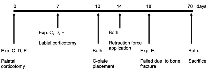
Fig. 3 Schematic illustration of the experimental design. Exp., Experimental group; Both., control group (Group I) and experimental group (Group II).
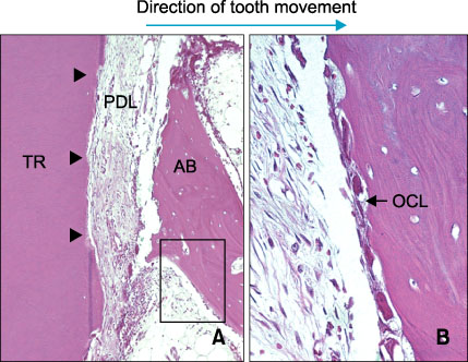
Fig. 4 Microphotographs of periodontal tissue on a labio-lingual section of the retracted maxillary anterior tooth (hematoxylin and eosin (H&E) stain) for lingual area (pressure side) for group I (dog A). A, Periodontal ligamental tissue was lost and root cementum was also partly absent. Note the demarcation line between root dentin and cementum was partially lost (H&E, × 100). Black framed area is magnified in B. Short black arrow heads show root resorption; B, osteoclasts were observed on the surface of resorbing alveolar bone (H&E, × 400). AB, Alveolar bone; PDL, periodontal ligament; TR, tooth root; OCL, osteoclasts.
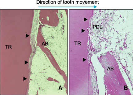
Fig. 5 Microphotographs of periodontal tissue on the lingual area (pressure side) for group I (dog B). A, Odontoclasts were observed on the surface of the resorbing tooth surface. Attachment loss of periodontal ligament and loss of cementum continuity are seen (H&E, × 40); B, higher magnification view of compression side (H&E, × 100). Short black arrow heads show root resorption. AB, Alveolar bone; PDL, periodontal ligament; TR, tooth root.
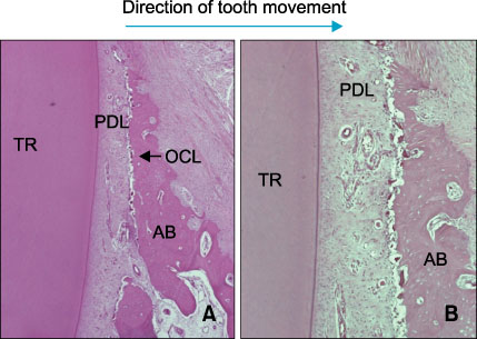
Fig. 6 Microphotographs of periodontal tissue on the lingual area (pressure side) for group II (dog C). A, Many osteoclasts are observed on the surface of alveolar bone, but no root resorption is observed. Note the intact continuous cementum line of the dental root (H&E, × 40); B, higher magnification view of the compression side (H&E, × 100). AB, Alveolar bone; PDL, periodontal ligament; TR, tooth root; OCL, osteoclasts.
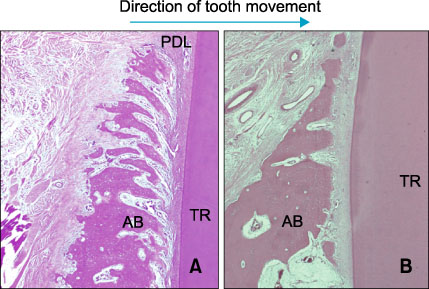
Fig. 7 Microphotographs of periodontal tissue on the labial area (tension side) for group I (dog B) and group II (dog C). A, Many osteoblasts are observed on the surface of alveolar bone for group I (dog B). There are many capillaries in the marrow space of new bone formation, however, resorption activities of bone or root were not observed. Note that the newly formed bone trabeculae were directed horizontally coinciding with the direction of tooth movement (H&E, × 40); B, histologic features of group II (dog C) were similar to Group I but with less extent of new bone formation (H&E, × 40). AB, Alveolar bone; PDL, periodontal ligament; TR, tooth root.
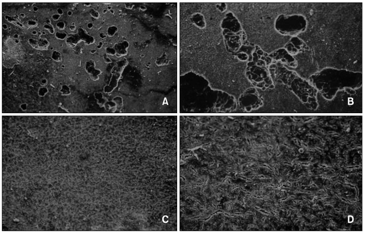
Fig. 8 Scanning electron microscopy images of the dental root surface from group I (dogs A and B) and group II (dogs C and D). A, Image of group I (dog A) shows resorption of cementum. Many resorption lacunae were clustered together and the sizes of lacunae were similar although the depths varied (SEM, × 100); B, higher magnification image of group I (dog A) shows round resorption lacunae with definite boundaries. Several small holes were visible in the bottom of some lacunae which are considered to be dentinal tubules (SEM, × 300); C, image of group II (dog D) shows smooth root surfaces without resorption lacunae (SEM, × 100); D, higher magnification of image of group II (dog D) at SEM, × 300.
Cited by 1 articles
-
Torque control during lingual anterior retraction without posterior appliances
Sung-Seo Mo, Seong-Hun Kim, Sang-Jin Sung, Kyu-Rhim Chung, Yun-Sic Chun, Yoon-Ah Kook, Gerald Nelson
Korean J Orthod. 2013;43(1):3-14. doi: 10.4041/kjod.2013.43.1.3.
Reference
-
1. Melsen B. Melsen B, editor. Limitations in adult orthodontics. Current controversies in orthodontics. 1991. 1st ed. Hanover Park, IL: Quintessence Publishing Co, Inc.;147–180.2. Vardimon AD, Oren E, Ben-Bassat Y. Cortical bone remodeling/tooth movement ratio during maxillary incisor retraction with tip versus torque movements. Am J Orthod Dentofacial Orthop. 1998. 114:520–529.
Article3. Bojrab DG, Dumas JE, Lahrman DE. JCO/interviews Dr. David G. Bojrab, Dr. James E. Dumas, Dr. Don E. Lahrman on surgical-orthodontics. J Clin Orthod. 1977. 11:330–342.4. Wilcko WM, Wilcko T, Bouquot JE, Ferguson DJ. Rapid orthodontics with alveolar reshaping: two case reports of decrowding. Int J Periodontics Restorative Dent. 2001. 21:9–19.5. Chen YR, Yeow VK. Multiple-segment osteotomy in maxillofacial surgery. Plast Reconstr Surg. 1999. 104:381–388.
Article6. Kole H. Surgical operations on the alveolar ridge to correct occlusal abnormalities. Oral Surg Oral Med Oral Pathol. 1959. 12:413–420.
Article7. Wilcko MT, Wilcko WM, Pulver JJ, Bissada NF, Bouquot JE. Accelerated osteogenic orthodontics technique: a 1-stage surgically facilitated rapid orthodontic technique with alveolar augmentation. J Oral Maxillofac Surg. 2009. 67:2149–2159.
Article8. Wilcko MT, Wilcko WM, Murphy KG, Carroll WJ, Ferguson DJ, Miley DD, et al. Full-thickness flap/subepithelial connective tissue grafting with intramarrow penetrations: three case reports of lingual root coverage. Int J Periodontics Restorative Dent. 2005. 25:561–569.9. Suya H. Hosl E, Baldauf A, editors. Corticotomy in orthodontics. Mechanical and biological basics in orthodontic therapy. 1991. Heidelberg, Germany: Huthig Buch;207–226.10. Lee BS, Hwang HW, Chung KR. Clinical use of corticotomies in adult orthodontics. J Korean Assoc Maxillofac Plast Reconstr Surg. 1999. 21:303–311.11. Chung KR, Oh MY, Ko SJ. Corticotomy-assisted orthodontics. J Clin Orthod. 2001. 35:331–339.12. Chung KR, Mitsugi M, Lee BS, Kanno T, Lee W, Kim SH. Speedy surgical orthodontic treatment with skeletal anchorage in adults--sagittal correction and open bite correction. J Oral Maxillofac Surg. 2009. 67:2130–2148.
Article13. Kim S, Park Y, Chung K. Severe anterior open bite malocclusion with multiple odontoma treated by C-lingual retractor and horseshoe mechanics. Angle Orthod. 2003. 73:206–212.14. Kim SH, Lee KB, Chung KR, Nelson G, Kim TW. Severe bimaxillary protrusion with adult periodontitis treated by corticotomy and compression osteogenesis. Korean J Orthod. 2009. 39:54–65.
Article15. Frost HM. The biology of fracture healing. An overview for clinicians. Part I. Clin Orthop Relat Res. 1989. (248):283–293.
Article16. Frost HM. The biology of fracture healing. An overview for clinicians. Part II. Clin Orthop Relat Res. 1989. (248):294–309.
Article17. Gwack C, Kim SS, Park SB, Son WS, Kim YD, Jun ES, et al. The expression of MMP-1, -8, and -13 mRNA in the periodontal ligament of rats during tooth movement with cortical punching. Korean J Orthod. 2008. 38:187–201.
Article18. Park WK, Kim SS, Park SB, Son WS, Kim YD, Jun ES, et al. The effect of cortical punching on the expression of OPG, RANK, and RANKL in the periodontal tissue during tooth movement in rats. Korean J Orthod. 2008. 38:159–174.
Article19. Chung KR, Kim SH, Lee BS. Speedy surgical-orthodontic treatment with temporary anchorage devices as an alternative to orthognathic surgery. Am J Orthod Dentofacial Orthop. 2009. 135:787–798.
Article20. Kim DH, Park YG, Kang SG. The effects of electrical current from a micro-electrical device on tooth movement. Korean J Orthod. 2008. 38:337–346.
Article21. Yoshikawa Y, Deguchi T, Eda S. Pulpal and radicular changes following maxillary subapical corticotomy. Endod Dent Traumatol. 1992. 8:245–247.
Article22. Lee JK, Chung KR, Baek SH. Treatment outcomes of orthodontic treatment, corticotomy-assisted orthodontic treatment, and anterior segmental osteotomy for bimaxillary dentoalveolar protrusion. Plast Reconstr Surg. 2007. 120:1027–1036.
Article23. Iino S, Sakoda S, Ito G, Nishimori T, Ikeda T, Miyawaki S. Acceleration of orthodontic tooth movement by alveolar corticotomy in the dog. Am J Orthod Dentofacial Orthop. 2007. 131:448.e1–448.e8.
Article24. Wang L, Lee W, Lei DL, Liu YP, Yamashita DD, Yen SL. Tisssue responses in corticotomy- and osteotomy-assisted tooth movements in rats: histology and immunostaining. Am J Orthod Dentofacial Orthop. 2009. 136:770.e1–770.e11.
Article25. Chung KR, Kook YA, Kim SH, Mo SS, Jung JA. Class II malocclusion treated by combining a lingual retractor and a palatal plate. Am J Orthod Dentofacial Orthop. 2008. 133:112–123.
Article
- Full Text Links
-
- Actions
-
Cited
- CITED
-
- Close
- Share
-
- Similar articles
-
- Severe bimaxillary protrusion with adult periodontitis treated by corticotomy and compression osteogenesis
- The endocannabinoid system in zebrafish and its potential to study the effects of Cannabis in humans
- An effect of immediate orthodontic force on palatal endosseous appliance(C-Palatal PlateTM) in beagle Dog
- Surgical Dilatational Tracheostomy to Prevent Post-Tracheostomy Tracheal Stenosis: Preliminary Results in a Growing Animal Model
- Corticotomy and the molar uprighting
