New three-dimensional cephalometric analyses among adults with a skeletal Class I pattern and normal occlusion
- Affiliations
-
- 1Graduate School, The Catholic University of Korea, Seoul, Korea.
- 2Postgraduate Orthodontic Program, Arizona School of Dentistry and Oral Health, A.T. Still University, Mesa, AZ, USA.
- 3Graduate School of Dentistry, Kyung Hee University, Seoul, Korea.
- 4Department of Orthodontics, Seoul St. Mary's Hospital, The Catholic University of Korea, Seoul, Korea. kook190036@yahoo.com
- KMID: 1975143
- DOI: http://doi.org/10.4041/kjod.2013.43.2.62
Abstract
OBJECTIVE
The purpose of this study was to assess new three-dimensional (3D) cephalometric variables, and to evaluate the relationships among skeletal and dentoalveolar variables through 3D cephalometric analysis.
METHODS
Cone-beam computed tomography (CBCT) scans were acquired from 38 young adults (18 men and 20 women; 22.6 +/- 3.2 years) with normal occlusion. Thirty-five landmarks were digitized on the 3D-rendered views. Several measurements were obtained for selected landmarks. Correlations among different variables were calculated by means of Pearson's correlation coefficient values.
RESULTS
The body of the mandible had a longer curve length in men (102.3 +/- 4.4 mm) than in women (94.5 +/- 4.7 mm) (p < 0.001), but there was no significant difference in the maxillary basal curve length. Men had significantly larger facial dimensions, whereas women had a larger gonial angle (117.0 +/- 4.0 vs. 113.8 +/- 3.3; p < 0.001). Strong-to-moderate correlation values were found among the vertical and transverse variables (r = 0.71 to 0.51).
CONCLUSIONS
The normative values of new 3D cephalometric parameters, including the maxillary and mandibular curve length, were obtained. Strong-to-moderate correlation values were found among several vertical and transverse variables through 3D cephalometric analysis. This method of cephalometric analyses can be useful in diagnosis and treatment planning for patients with dentofacial deformities.
Keyword
MeSH Terms
Figure
-
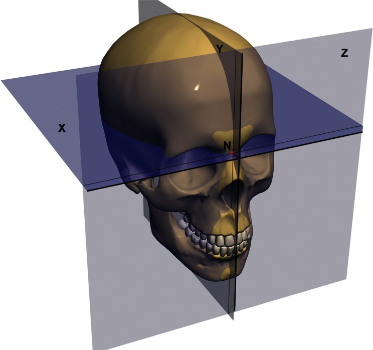
Figure 1 Reorientation of head and coordinate system. N, Nasion; X, the horizontal plane; Y, the midsagittal plane; Z, the vertical plane.
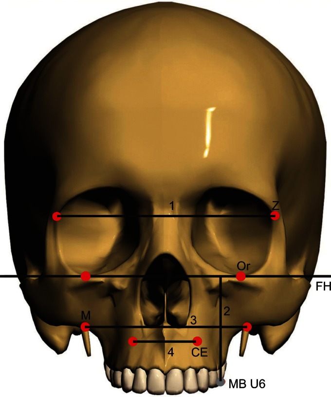
Figure 2 Frontal view shows maxillary landmarks and variables: Z, Zygomatic point; Or, orbitale; FH, Frankfort horizontal plane; M, maxillary point; CE, canine eminence; MB U6, mesiobuccal cusp of upper 1st molar; 1, upper facial width; 2, maxillary height; 3, posterior maxillary basal width; 4, anterior maxillary basal width.
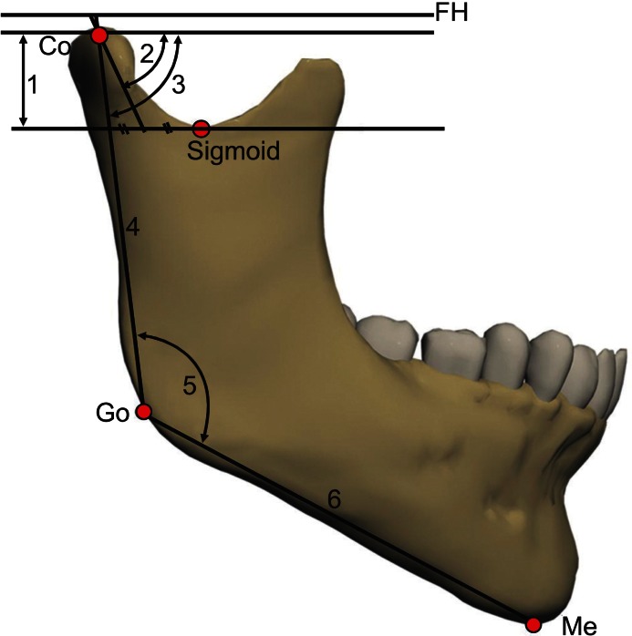
Figure 3 Lateral view of the mandible shows ramal and condylar landmarks and parameters: FH, Frankfort horizontal plane; Co, condylion; Go, gonion; Me, menton; 1, condylar height; 2, condylar anteroposterior inclination; 3, ramal anteroposterior inclination; 4, ramal length; 5, gonial angle; 6, mandibular body length.
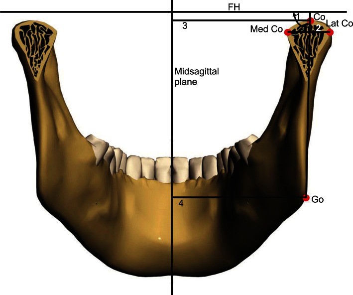
Figure 4 Posterior view of the mandible shows ramal and condylar landmarks and parameters: FH, Frankfort horizontal plane; Co, condylion; Med Co, medial condyle; Lat Co, lateral condyle; Go, gonion; 1, ramal mediolateral inclination; 2, condylar width; 3, condyle to midsagittal plane; 4, gonion to midsagittal plane.
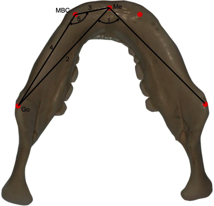
Figure 5 Mandibular body variables: Me, menton; MBC, mandibular body curve; Go, gonion; 1, menton angle; 2, mandibular body length; 3, anterior mandibular body length; 4, posterior mandibular body length; 5, MBC angle.
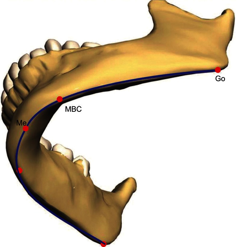
Figure 6 Mandibular basal curve length: Go, gonion; Me, menton; MBC, mandibular body curve.
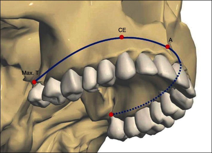
Figure 7 Maxillary basal curve length: Max. T, maxillary tuberosity; CE, canine eminence; A, A point.
Cited by 4 articles
-
The genial tubercle: A prospective novel landmark for the diagnosis of mandibular asymmetry
Seung-Youp Lee, Dong-Soon Choi, Insan Jang, Geun-Su Song, Bong-Kuen Cha
Korean J Orthod. 2017;47(1):50-58. doi: 10.4041/kjod.2017.47.1.50.Cone-beam computed tomography analysis of transverse dental compensation in patients with skeletal Class III malocclusion and facial asymmetry
Ji-Yea Lee, Sung-Hoon Han, Hyeong-Seok Ryu, Hee-Min Lee, Sang-Cheol Kim
Korean J Orthod. 2018;48(6):357-366. doi: 10.4041/kjod.2018.48.6.357.Diagnostic methods for assessing maxillary skeletal and dental transverse deficiencies: A systematic review
Dena Sawchuk, Kris Currie, Manuel Lagravere Vich, Juan Martin Palomo, Carlos Flores-Mir
Korean J Orthod. 2016;46(5):331-342. doi: 10.4041/kjod.2016.46.5.331.A ramus cortical bone harvesting technique without bone marrow invasion
Jeong-Kui Ku, Min-Soo Ghim, Jung Ho Park, Dae Ho Leem
J Korean Assoc Oral Maxillofac Surg. 2023;49(2):100-104. doi: 10.5125/jkaoms.2023.49.2.100.
Reference
-
1. Baumrind S, Frantz RC. The reliability of head film measurements. 1. Landmark identification. Am J Orthod. 1971; 60:111–127. PMID: 5283996.2. Ahlqvist J, Eliasson S, Welander U. The cephalometric projection. Part II. Principles of image distortion in cephalography. Dentomaxillofac Radiol. 1983; 12:101–108. PMID: 6584358.3. Kusnoto B, Evans CA, BeGole EA, de Rijk W. Assessment of 3-dimensional computer-generated cephalometric measurements. Am J Orthod Dentofacial Orthop. 1999; 116:390–399. PMID: 10511666.
Article4. Rousset MM, Simonek F, Dubus JP. A method for correction of radiographic errors in serial three-dimensional cephalometry. Dentomaxillofac Radiol. 2003; 32:50–59. PMID: 12820854.
Article5. Nakasima A, Terajima M, Mori N, Hoshino Y, Tokumori K, Aoki Y, et al. Three-dimensional computer-generated head model reconstructed from cephalograms, facial photographs, and dental cast models. Am J Orthod Dentofacial Orthop. 2005; 127:282–292. PMID: 15775942.
Article6. Ludlow JB, Gubler M, Cevidanes L, Mol A. Precision of cephalometric landmark identification: conebeam computed tomography vs conventional cephalometric views. Am J Orthod Dentofacial Orthop. 2009; 136:312.e1–312.e10. PMID: 19732656.
Article7. Ludlow JB, Laster WS, See M, Bailey LJ, Hershey HG. Accuracy of measurements of mandibular anatomy in cone beam computed tomography images. Oral Surg Oral Med Oral Pathol Oral Radiol Endod. 2007; 103:534–542. PMID: 17395068.
Article8. Cevidanes LH, Bailey LJ, Tucker GR Jr, Styner MA, Mol A, Phillips CL, et al. Superimposition of 3D cone-beam CT models of orthognathic surgery patients. Dentomaxillofac Radiol. 2005; 34:369–375. PMID: 16227481.
Article9. Cevidanes LH, Bailey LJ, Tucker SF, Styner MA, Mol A, Phillips CL, et al. Three-dimensional cone-beam computed tomography for assessment of mandibular changes after orthognathic surgery. Am J Orthod Dentofacial Orthop. 2007; 131:44–50. PMID: 17208105.
Article10. van Vlijmen OJ, Maal TJ, Bergé SJ, Bronkhorst EM, Katsaros C, Kuijpers-Jagtman AM. A comparison between two-dimensional and three-dimensional cephalometry on frontal radiographs and on cone beam computed tomography scans of human skulls. Eur J Oral Sci. 2009; 117:300–305. PMID: 19583759.
Article11. van Vlijmen OJ, Bergé SJ, Bronkhorst EM, Swennen GR, Katsaros C, Kuijpers-Jagtman AM. A comparison of frontal radiographs obtained from cone beam CT scans and conventional frontal radiographs of human skulls. Int J Oral Maxillofac Surg. 2009; 38:773–778. PMID: 19369033.
Article12. Gribel BF, Gribel MN, Manzi FR, Brooks SL, McNamara JA Jr. From 2D to 3D: an algorithm to derive normal values for 3-dimensional computerized assessment. Angle Orthod. 2011; 81:3–10. PMID: 20936948.
Article13. Farronato G, Garagiola U, Dominici A, Periti G, de Nardi S, Carletti V, et al. "Ten-point" 3D cephalometric analysis using low-dosage cone beam computed tomography. Prog Orthod. 2010; 11:2–12. PMID: 20529623.
Article14. Cheung LK, Chan YM, Jayaratne YS, Lo J. Three-dimensional cephalometric norms of Chinese adults in Hong Kong with balanced facial profile. Oral Surg Oral Med Oral Pathol Oral Radiol Endod. 2011; 112:e56–e73. PMID: 21665497.
Article15. Ratner B. Statistical modeling and analysis for database marketing: effective techniques for mining big data. 2003. Boca Raton: Chapman and Hall/CRC;p. 17.16. Lee H, Bayome M, Kim SH, Kim KB, Behrents RG, Kook YA. Mandibular dimensions of subjects with asymmetric skeletal Class III malocclusion and normal occlusion compared with cone-beam computed tomography. Am J Orthod Dentofacial Orthop. 2012; 142:179–185. PMID: 22858326.
Article17. Lee CT. Standards for Korean adult facial relationships by various roentgeno-cephalometric analysis. Korean J Orthod. 1988; 18:459–474.18. You KH, Lee KJ, Lee SH, Baik HS. Three-dimensional computed tomography analysis of mandibular morphology in patients with facial asymmetry and man dibular prognathism. Am J Orthod Dentofacial Orthop. 2010; 138:540. PMID: 21055584.19. Huntjens E, Kiss G, Wouters C, Carels C. Condylar asymmetry in children with juvenile idiopathic arthritis assessed by cone-beam computed tomography. Eur J Orthod. 2008; 30:545–551. PMID: 19054811.
Article20. Enlow DH, Hans MG. Essentials of facial growth. 1996. Philadelphia: W.B. Saunders;p. 72.21. Kim EJ, Palomo JM, Kim SS, Lim HJ, Lee KM, Hwang HS. Maxillofacial characteristics affecting chin deviation between mandibular retrusion and prognathism patients. Angle Orthod. 2011; 81:988–993. PMID: 21649508.
Article22. Shah SM, Joshi MR. An assessment of asymmetry in the normal craniofacial complex. Angle Orthod. 1978; 48:141–148. PMID: 277077.
- Full Text Links
-
- Actions
-
Cited
- CITED
-
- Close
- Share
-
- Similar articles
-
- Posteroanterior cephalometric characteristics in skeletal Class III malocclusion
- A comparative study on chewing movement in normal occlusion and skeletal Class III malocclusion
- A Lateral Cephalometric Study of Maxillofacial Morphologic Features in Class III Malocclusion Children
- A study on the difference of the skeletal maturity in normal occlusion and malocclusion
- A roentgenocephalomtric study on maxillary protrusion
