
Bilateral postoperative maxillary cysts after orthognathic surgery: A case report
- Affiliations
-
- 1Department of Oral and Maxillofacial Radiology and Dental Research Institute, School of Dentistry, Seoul National University, Seoul, Korea. raychoi@snu.ac.kr
- KMID: 1974502
- DOI: http://doi.org/10.5624/isd.2014.44.4.321
Abstract
- Postoperative maxillary cysts are locally aggressive lesions, usually developing as delayed complications many years after radical antral surgery. This report describes a case of bilateral postoperative maxillary cysts following orthognathic surgery performed approximately 21 years previously. The patient complained of stinging pain on her right cheek. Radiographic examination revealed low-attenuation lesions on both maxillary sinuses with discontinuously corticated margins without distinct expansion or bone destruction. The cysts were enucleated with the removal of metal plates and screws for pain relief. Histopathological examination confirmed the diagnosis of postoperative maxillary cysts lined by ciliated, pseudostratified columnar cells. The patient has remained asymptomatic thus far, and there was no evidence of local recurrence at 21 months of postoperative follow-up.
Keyword
MeSH Terms
Figure
-
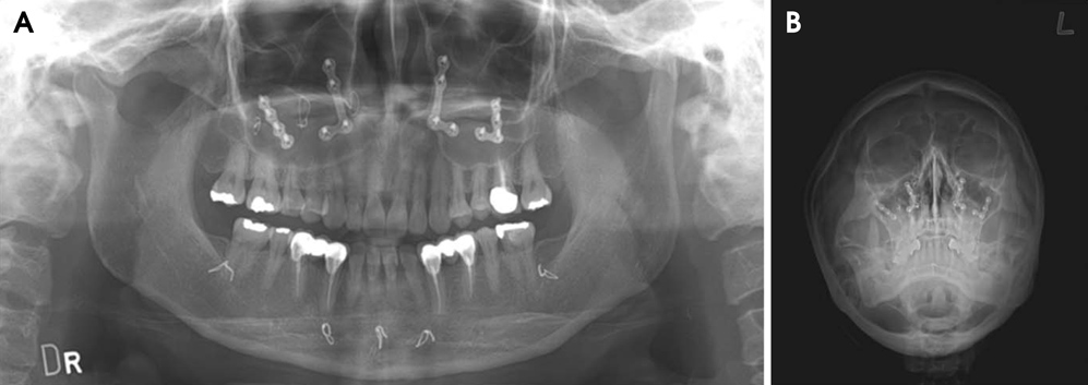
Fig. 1 A. Panoramic radiograph shows dome-shaped homogeneous radiopacities at the lower regions in both maxillary sinuses. There are metal plates, screws in the maxilla, and wires on the mandible from the previous orthognathic surgery. B. Waters' radiograph shows increased haziness at the lower regions in both maxillary sinuses, but there is no distinct expansion or bone destruction.

Fig. 2 Computed tomography images show low-attenuation lesions in both maxillary sinuses. A. Axial image reveals no distinct expansion and bone destruction of the lesions. Postoperative signs such as antral wall thickening, sclerotic change, and loss of continuity of the cortical antral wall lining are not seen. B. Coronal image shows that the lesions extended slightly below the floor of both nasal sinuses. C and D. Sagittal images of right (C) and left (D) maxillary sinuses reveal the undulated margins, which were discontinuously corticated.
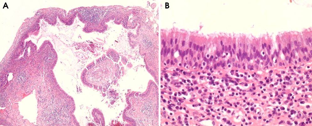
Fig. 3 A. Histopathological examination shows the inflammatory cells infiltrating around the cystic cavity (H&E stain, 40×). B. The cyst is lined by ciliated, pseudostratified columnar cells (H&E stain, 400×).
Reference
-
1. Hasegawa M, Saito Y, Watanabe I, Kern EB. Postoperative mucoceles of the maxillary sinus. Rhinology. 1979; 17:253–256.2. Pe MB, Sano K, Kitamura A, Inokuchi T. Computed tomography in the evaluation of postoperative maxillary cysts. J Oral Maxillofac Surg. 1990; 48:679–684.
Article3. Lee GI, Park TW. Clinical and radiological study of the postoperative maxillary cyst. J Korean Acad Oral Maxillofac Radiol. 1994; 24:47–55.4. Choi KS. The post-operative maxillary cyst: report of 14 cases. J Korean Dent Assoc. 1989; 27:1049–1057.5. Sugar AW, Walker DM, Bounds GA. Surgical ciliated (postoperative maxillary) cysts following mid-face osteotomies. Br J Oral Maxillofac Surg. 1990; 28:264–267.
Article6. Hayhurst DL, Moenning JE, Summerlin DJ, Bussard DA. Surgical ciliated cyst: a delayed complication in a case of maxillary orthognathic surgery. J Oral Maxillofac Surg. 1993; 51:705–709.
Article7. Amin M, Witherow H, Lee R, Blenkinsopp P. Surgical ciliated cyst after maxillary orthognathic surgery: report of a case. J Oral Maxillofac Surg. 2003; 61:138–141.
Article8. Bourgeois SL Jr, Nelson BL. Surgical ciliated cyst of the mandible secondary to simultaneous Le Fort I osteotomy and genioplasty: report of case and review of the literature. Oral Surg Oral Med Oral Pathol Oral Radiol Endod. 2005; 100:36–39.
Article9. Heo MS, Song MY, Lee SS, Choi SC, Park TW. A comparative study of the radiological diagnosis of postoperative maxillary cyst. Dentomaxillofac Radiol. 2000; 29:347–351.
Article10. Noyek AM. Radiology of the maxillary sinus after Caldwell-Luc surgery. Otolaryngol Clin North Am. 1976; 9:135–151.
Article11. Unger JM, Dennison BF, Duncavage JA, Toohill RJ. The radiological appearance of the post-Caldwell-Luc maxillary sinus. Clin Radiol. 1986; 37:77–81.
Article12. Kaneshiro S, Nakajima T, Yoshikawa Y, Iwasaki H, Tokiwa N. The postoperative maxillary cyst: report of 71 cases. J Oral Surg. 1981; 39:191–198.13. Kim JM, Cho JH, Jung HV, Choi JM, Kim JS, Kim JK. Postoperative maxillary cyst after maxillary orthognathic surgery: report of an unusual case. J Rhinol. 2012; 19:60–62.14. Kim JK, Choi YS, Kim SY, Yi CK. Postoperative maxillary cyst after orthognathic surgery. J Korean Assoc Maxillofac Plast Reconstr Surg. 1996; 18:120–124.15. Lyon HE. Reliability of panoramic radiography in the diagnosis of maxillary sinus pathosis. Oral Surg Oral Med Oral Pathol. 1973; 35:124–128.
Article16. Ohba T, Katayama H. Comparison of panoramic radiography and Water's projection in the diagnosis of maxillary sinus disease. Oral Surg Oral Med Oral Pathol. 1976; 42:534–538.
Article17. Choi SC, Ahn HK. Radiologic study of the maxillary sinusitis. J Korean Acad Oral Maxillofac Radiol. 1981; 11:41–50.18. Cable HR, Jeans WD, Cullen RJ, Bull PD, Maw AR. Computerized tomography of the Caldwell-Luc cavity. J Laryngol Otol. 1981; 95:775–783.
Article
- Full Text Links
-
- Actions
-
Cited
- CITED
-
- Close
- Share
-
- Similar articles
-
- Surgical ciliated cyst of the mandible after orthognathic surgery: a case report with review of the literature
- Post-Operative Maxillary Cyst after Maxillary Orthognathic Surgery: Report of an Unusual Case
- Massive Postoperative Bleeding: A Case Report
- Removal of fungus ball of the right maxillary sinus via LeFort I osteotomy: A case report
- Treatment in Bimaxillary Prognathism with Anterior Open Bite: A Case Report


