Glandular odontogenic cyst mimicking ameloblastoma in a 78-year-old female: A case report
- Affiliations
-
- 1Department of Oral and Maxillofacial Radiology and Wonkwang Dental Research Institute, College of Dentistry, Wonkwang University, Iksan, Korea. eebydo@wonkwang.ac.kr
- 2Department of Oral and Maxillofacial Surgery, College of Dentistry, Wonkwang University, Iksan, Korea.
- 3Department of Oral and Maxillofacial Pathology, College of Dentistry, Daejeon Dental Hospital, Wonkwang University, Daejeon, Korea.
- KMID: 1974490
- DOI: http://doi.org/10.5624/isd.2014.44.3.249
Abstract
- Glandular odontogenic cyst (GOC) is a rare, potentially aggressive jaw lesion. The common radiographic features include a well-defined radiolucency with distinct borders, presenting a uni- or multilocular appearance. A cystic lesion in the posterior mandible of a 78-year-old female was incidentally found. Radiographs showed a unilocular lesion with a scalloped margin, external root resorption of the adjacent tooth, and cortical perforation. This lesion had changed from a small ovoid shape to a more expanded lesion in a period of four years. The small lesion showed unilocularity with a smooth margin and a well-defined border, but the expanded lesion produced cortical perforation and a lobulated margin. The provisional diagnosis was an ameloblastoma, whereas the histopathological examination revealed a GOC. This was a quite rare case, given that this radiographic change was observed in the posterior mandible of an elderly female. This case showed that a GOC can grow even in people in their seventies, changing from the unilocular form to an expanded, lobulated lesion. Here, we report a case of GOC with characteristic radiographic features.
Keyword
MeSH Terms
Figure
-
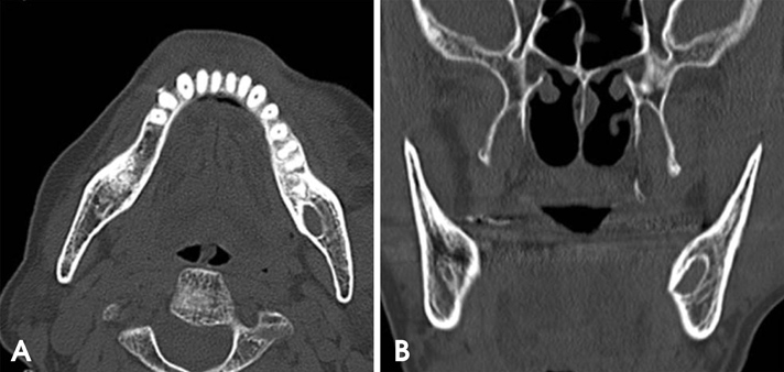
Fig. 1 Axial (A) and coronal (B) computed tomography images show an ovoid, unilocular cystic lesion on the left mandibular angle.
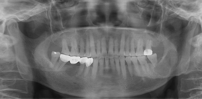
Fig. 2 The panoramic radiograph shows an ovoid, lobulated lesion with a well-defined margin from the left lower second molar to the left mandibular ramus. The external root resorption of the second molar is remarkable.

Fig. 3 The cone-beam computed tomographic images represent a unilocular lesion with a scalloped margin, cortical thinning, perforation, and erosion. There was also mild expansion of the lingual cortex.

Fig. 4 Photomicrograph shows multiple cystic compartments (H&E stain, 100×).
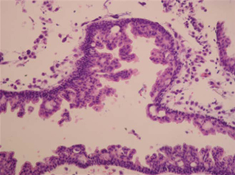
Fig. 5 The cyst is lined by several surface eosinophilic cuboidal cells. Microcysts and papillary projections formed adjacent to "open" microcysts (H&E stain, 200×).
Reference
-
1. Hussain K, Edmondson HD, Browne RM. Glandular odontogenic cysts. Diagnosis and treatment. Oral Surg Oral Med Oral Pathol Oral Radiol Endod. 1995; 79:593–602.2. Kaplan I, Anavi Y, Hirshberg A. Glandular odontogenic cyst: a challenge in diagnosis and treatment. Oral Dis. 2008; 14:575–581.
Article3. Macdonald-Jankowski DS. Glandular odontogenic cyst: systematic review. Dentomaxillofac Radiol. 2010; 39:127–139.
Article4. Kramer IR, Pindborg JJ, Shear M. Histological typing of odontogenic tumors. 2nd ed. Berlin/New York: Springer Verlag;1992.5. Padayachee A, Van Wyk CW. Two cystic lesions with features of both the botryoid odontogenic cyst and the central mucoepidermoid tumour: sialo-odontogenic cyst? J Oral Pathol. 1987; 16:499–504.
Article6. Gardner DG, Kessler HP, Morency R, Schaffner DL. The glandular odontogenic cyst: an apparent entity. J Oral Pathol. 1988; 17:359–366.
Article7. Manor R, Anavi Y, Kaplan I, Calderon S. Radiological features of glandular odontogenic cyst. Dentomaxillofac Radiol. 2003; 32:73–79.
Article8. Tambawala SS, Karjodkar FR, Yadav A, Sansare K, Sontakke S. Glandular odontogenic cyst: a case report. Imaging Sci Dent. 2014; 44:75–79.
Article9. Ramer M, Montazem A, Lane SL, Lumerman H. Glandular odontogenic cyst: report of a case and review of the literature. Oral Surg Oral Med Oral Pathol Oral Radiol Endod. 1997; 84:54–57.10. Koppang HS, Johannessen S, Haugen LK, Haanaes HR, Solheim T, Donath K. Glandular odontogenic cyst (sialo-odontogenic cyst): report of two cases and literature review of 45 previously reported cases. J Oral Pathol Med. 1998; 27:455–462.
Article11. Noffke C, Raubenheimer EJ. The glandular odontogenic cyst: clinical and radiological features; review of the literature and report of nine cases. Dentomaxillofac Radiol. 2002; 31:333–338.
Article12. White SC, Pharoah MJ. Oral radiology: principles and interpretation. 4th ed. St. Louis: Mosby;1999. p. 364.13. Kaplan I, Anavi Y, Manor R, Sulkes J, Calderon S. The use of molecular markers as an aid in the diagnosis of glandular odontogenic cyst. Oral Oncol. 2005; 41:895–902.
Article14. Fowler CB, Brannon RB, Kessler HP, Castle JT, Kahn MA. Glandular odontogenic cyst: analysis of 46 cases with special emphasis on microscopic criteria for diagnosis. Head Neck Pathol. 2011; 5:364–375.
Article15. Shear MS, Speight PM. Cysts of the oral and maxillofacial regions. 4th ed. Oxford: Blackwell Pub;2007. p. 94–99.16. Boffano P, Cassarino E, Zavattero E, Campisi P, Garzino-Demo P. Surgical treatment of glandular odontogenic cysts. J Craniofac Surg. 2010; 21:776–780.
Article
- Full Text Links
-
- Actions
-
Cited
- CITED
-
- Close
- Share
-
- Similar articles
-
- Re: Byung-Do Lee, Wan Lee, Kyung-Hwan Kwon, Moon-Ki Choi, Eun-Joo Choi and Jung-Hoon Yoon. Glandular odontogenic cyst mimicking ameloblastoma in a 78-year-old female: A case report. Imaging Science in Dentistry 2014; 44(3): 249-52.
- Response to letter to the editors "Re: Byung-Do Lee, Wan Lee, Kyung-Hwan Kwon, Moon-Ki Choi, Eun-Joo Choi and Jung-Hoon Yoon. Glandular odontogenic cyst mimicking ameloblastoma in a 78-year-old female: a case report. Imaging Science in Dentistry 2014; 44(3): 249-52."
- Glandular odontogenic cyst of mandible: case report
- Unicystic ameloblastoma arising from dentigerous cyst: case report and literature review
- Ameloblastoma originated from a dentigerous cyst: a case report
