Multiple fibromyxomas of the jaws: A case report
- Affiliations
-
- 1Oral Radiology Department, Faculty of Oral and Dental Medicine, Cairo University, Cairo, Egypt. m_khalifa_z@yahoo.com
- KMID: 1974488
- DOI: http://doi.org/10.5624/isd.2014.44.3.237
Abstract
- Fibromyxoma of the jaw is a rare benign mesenchymal odontogenic tumor with locally aggressive behavior. In the present report, a 13-year-old female patient presented to our university hospital with delayed eruption of some of her teeth. A panoramic radiograph taken at the initial examination revealed four pericoronal radiolucencies related to the four third molars. Thereafter, a magnetic resonance imaging (MRI) examination was performed. After the surgical removal of these molars, the microscopic examination diagnosed the four lesions as fibromyxomas. Here, we have discussed the clinical, panoramic radiography, MRI, and histopathological findings of the case.
Keyword
MeSH Terms
Figure
-
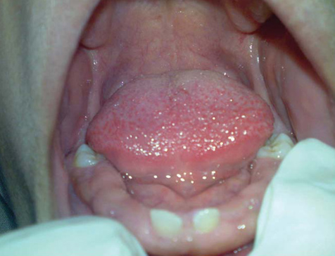
Fig. 1 Intra-oral photograph shows absence of multiple teeth in the mandible.
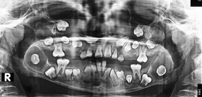
Fig. 2 Panoramic radiograph shows four pericoronal radiolucencies related to unerupted third molars as well as multiple impacted teeth with delayed development.
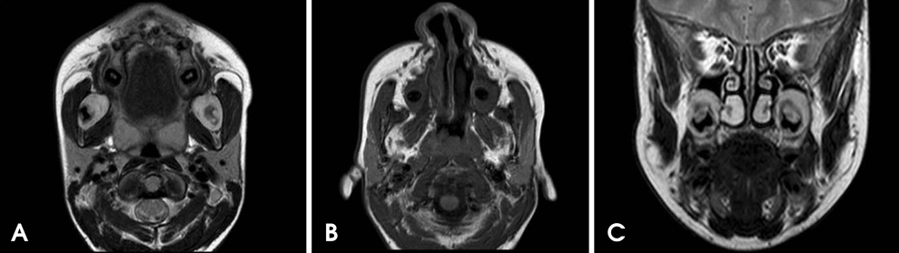
Fig. 3 A. Axial T2-weighted magnetic resonance (MR) image shows bilateral pericoronal lesions with a slightly hyper-intense heterogeneous signal and a black void representing the impacted tooth on the right side as well as the buccal and lingual expansion of the ramus cortices. B. Axial T1-weighted MR image shows bilateral pericoronal lesions with an intermediate signal invaginating the maxillary sinus. C. Coronal T2-weighted MR image shows bilateral pericoronal lesions with a slightly hyper-intense heterogeneous signal with the invagination of both maxillary sinuses.
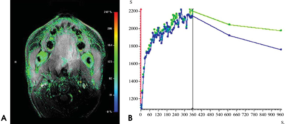
Fig. 4 A. Axial colored relative enhancement map of magnetic resonance imaging (MRI) shows a strong heterogeneous enhancement of the mandibular lesions. B. Time-intensity curves of both mandibular lesions show the same behavior: a rapid initial rise in the first few seconds that continues at a relatively slow rate to reach its maximum enhancement at 360 s. Then, it shows a gradual washout in the delayed phase (10 min) and another at a relatively slow rate in the super-delayed phase (15 min).
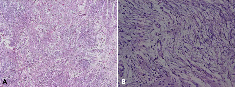
Fig. 5 Microphotographs show (A) a hypocellular tumor with a focally collagenous background (hematoxylin and eosin (H&E) stain, 10 ×) and (B) spindle and stellate cells with bland nuclear features arranged in a myxoid background (H&E stain, 40×).
Reference
-
1. Neville BW. Oral and maxillofacial pathology. 3rd ed. St. Louis: Saunders;2009.2. Sivakumar G, Kavitha B, Saraswathi T, Sivapathasundharam B. Odontogenic myxoma of maxilla. Indian J Dent Res. 2008; 19:62–65.
Article3. Shah A, Lone P, Latoo S, Ahmed I, Malik A, Hassan S, et al. Odontogenic myxoma of the maxilla: a report of a rare case and review on histogenetic and diagnostic concepts. Natl J Maxillofac Surg. 2011; 2:189–195.
Article4. Wawro NW, Reed J. Fibromyxoma of the mandible: report of two cases. Ann Surg. 1950; 132:1138–1143.5. Allen PS. Fibromyxoma of the mandible: case report and radiographic considerations. J Am Dent Assoc. 1980; 101:930–931.
Article6. Abiose BO, Ajagbe HA, Thomas O. Fibromyxomas of the jawbones - a study of ten cases. Br J Oral Maxillofac Surg. 1987; 25:415–421.7. Cervellini P, Crescenzi A, Di Zitti P. Mandibular fibromyxoma. Pathologica. 1993; 85:569–572.8. Piesold J, Meerbach W. Odontogenic fibromyxoma of the mandible. Mund Kiefer Gesichtschir. 1998; 2:44–47.9. Mishra A, Bhatia N, Shukla GK. Fibromyxoma maxilla. Indian J Otolaryngol Head Neck Surg. 2004; 56:293–295.
Article10. Berry S, Puri R. Fibromyxoma of the maxilla. Otolaryngol Head Neck Surg. 2006; 135:330–331.
Article11. Shahoon H, Esmaeili M, Nikhalat M, Farokhi E. Odontogenic fibromyxoma and odontogenic cyst in an eight year old boy: three-year follow-up. J Dent Res Dent Clin Dent Prospects. 2009; 3:103–105.12. Singaraju S, Wanjari SP, Parwani RN. Odontogenic myxoma of the maxilla: a report of a rare case and review of the literature. J Oral Maxillofac Pathol. 2010; 14:19–23.
Article13. Dietrich EM, Papaemmanouil S, Koloutsos G, Antoniades H, Antoniades K. Odontogenic fibromyxoma of the maxilla: a case report and review of the literature. Case Rep Med. 2011; 2011:238712.
Article14. Reddy GS, Kumar BS, Muppa R, Regonda SK, Tvs HK. Odontogenic fibromyxoma of maxilla: a rare case report. Case Rep Dent. 2013; 2013:345479.
Article15. Manne RK, Kumar VS, Venkata Sarath P, Anumula L, Mundlapudi S, Tanikonda R. Odontogenic myxoma of the mandible. Case Rep Dent. 2012; 2012:214704.
Article16. Mosqueda-Taylor A, Ledesma-Montes C, Caballero-Sandoval S, Portilla-Robertson J, Ruíz-Godoy Rivera LM, Meneses-García A. Odontogenic tumors in Mexico: a collaborative retrospective study of 349 cases. Oral Surg Oral Med Oral Pathol Oral Radiol Endod. 1997; 84:672–675.17. Martínez-Mata G, Mosqueda-Taylor A, Carlos-Bregni R, de Almeida OP, Contreras-Vidaurre E, Vargas PA, et al. Odontogenic myxoma: clinico-pathological, immunohistochemical and ultrastructural findings of a multicentric series. Oral Oncol. 2008; 44:601–607.
Article18. Keszler A, Dominguez FV, Giannunzio G. Myxoma in childhood: an analysis of 10 cases. J Oral Maxillofac Surg. 1995; 53:518–521.19. Raubenheimer EJ, Noffke CE. Central odontogenic fibromalike tumors, hypodontia, and enamel dysplasia: review of the literature and report of a case. Oral Surg Oral Med Oral Pathol Oral Radiol Endod. 2002; 94:74–77.
Article20. Mosqueda-Taylor A. New findings and controversies in odontogenic tumors. Med Oral Patol Oral Cir Bucal. 2008; 13:E555–E558.21. Sadeghi EM, Sewall SR, Dohse A, Novak TS. Odontogenic tumors that mimic a dentigerous cyst. Compend Contin Educ Dent. 1995; 16:500–504.22. Wood NK, Gauze PW. Differential diagnosis of oral and maxillofacial lesions. 5th ed. St. Louis: Mosby;1997. p. 279–294.23. Thunthy KH. Differential diagnosis of pericoronal radiolucencies with and without radiopacities. Gen Dent. 1999; 47:182–186.24. Miller CS, Bean LR. Pericoronal radiolucencies with and without radiopacities. Dent Clin North Am. 1994; 38:51–61.25. DelBalso AM, Hall RE. Advances in maxillofacial imaging. Curr Probl Diagn Radiol. 1993; 22:91–142.
Article26. Sumi Y, Miyaishi O, Ito K, Ueda M. Magnetic resonance imaging of myxoma in the mandible: a case report. Oral Surg Oral Med Oral Pathol Oral Radiol Endod. 2000; 90:671–676.
Article27. Asaumi J, Konouchi H, Hisatomi M, Kishi K. Odontogenic myxoma of maxillary sinus: CT and MR-pathologic correlation. Eur J Radiol. 2001; 37:1–4.
Article28. Aquilino RN, Tuji FM, Eid NL, Molina OF, Joo HY, Haiter Neto F. Odontogenic myxoma in the maxilla: a case report and characteristics on CT and MR. Oral Oncol Extra. 2006; 42:133–136.
Article29. Kawai T, Murakami S, Nishiyama H, Kishino M, Sakuda M, Fuchihata H. Diagnostic imaging for a case of maxillary myxoma with a review of the magnetic resonance images of myxoid lesions. Oral Surg Oral Med Oral Pathol Oral Radiol Endod. 1997; 84:449–454.
Article30. Asaumi J, Matsuzaki H, Hisatomi M, Konouchi H, Shigehara H, Kishi K. Application of dynamic MRI to differentiating odontogenic myxomas from ameloblastomas. Eur J Radiol. 2002; 43:37–41.
Article31. Feller L, Jadwat Y, Bouckaert M, Buskin A, Raubenheimer EJ. Enamel dysplasia with odontogenic fibroma-like hamartomas: review of the literature and report of a case. Oral Surg Oral Med Oral Pathol Oral Radiol Endod. 2006; 101:620–624.
Article32. Feller L, Wood NH, Anagnostopoulos C, Bouckaert M, Raubenheimer EJ, Kramer B, et al. Enamel dysplasia with hamartomatous atypical follicular hyperplasia: review of the literature and report of a case. SADJ. 2008; 63:96–97. 100–101.
- Full Text Links
-
- Actions
-
Cited
- CITED
-
- Close
- Share
-
- Similar articles
-
- Fibrous Dysplasia of the Jaws Associated with Secondary Hyperparathyroidism: A Case Report
- Multiple Ossifying-Fibroma In The Jaws: A Case Report
- Bilateral inflammatory cysts of the jaw: report of an unusual case
- Multiple fibro-osseous lesions of the jaws: A report of a rare case with a literature review
- Multidisciplinary approach for medication-related osteonecrosis of the jaws: a case report and literature review
