Diverse imaging characteristics of a mandibular intraosseous vascular lesion
- Affiliations
-
- 1Department of Oral Medicine and Radiology, Peoples Dental Academy, Bhopal, India. drsgin@gmail.com
- 2Department of Oral Pathology and Microbiology, Peoples Dental Academy, Bhopal, India.
- KMID: 1974476
- DOI: http://doi.org/10.5624/isd.2014.44.1.67
Abstract
- Intraosseous vascular lesions of the maxillofacial region are rare, and the differential diagnosis of intraosseous vascular malformations from other jaw lesions can be challenging. In the present case, magnetic resonance imaging and three-dimensional computed tomographic angiography (CTA) was used for diagnosis, and the lesion was treated wih surgical excision. Diverse characteristics such as the "honeycomb" and "sunburst" radiographic appearances and the absence of major peripheral feeder vessels in the CTA were noted. Intraosseous vascular malformations have a varied radiographic appearance, and the nomenclature of these lesions is equally diverse, with several overlapping terms. Pathologists do not generally differentiate among intraosseous vascular lesions on the basis of histopathology, although these lesions may present with contrasting immunohistochemical and clinical behaviors requiring varied treatment strategies. This case report highlights the need for multiple imaging modalities to differentiate among vascular lesions, as well as to better understand the behaviors of these unique lesions.
Keyword
MeSH Terms
Figure
-

Fig. 1 An intraoral photograph shows a dome shaped bluish alveolar swelling in the left mandibular premolar region.

Fig. 2 A. A panoramic radiograph shows a multilocular radiolucent lesion in the premolar region with small loculations giving a 'honeycomb appearance'. B. An occlusal radiograph shows a periosteal reaction as a "sun-burst" appearance. C. An intraoral periapical radiograph shows a multilocular radiolucent lesion in the premolar region with small loculations.

Fig. 3 Histopathologic examination shows frank blood aspirate with cytology showing plenty of red blood cells (H&E stain, 45×).
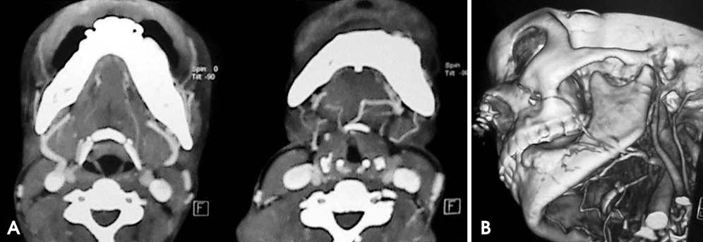
Fig. 4 A. CT images show trabeculated lesion in left hemi-mandible, with vascular lesion and feeder vessel seen supplying the lesion. B. A 3-dimensional CT angiogram shows a vascular lesion and feeder vessel.

Fig. 5 A. A coronal T1 weighted MR image shows an expansile low intensity lesion in marrow of left hemi-mandible. B. An axial T1 weighted MR image shows low signal intensity expansile lesion in marrow of left hemi-mandible with cortical break. C. An axial T2 weighted MR image shows a high signal expansile lesion in marrow of left hemi-mandible with cortical break.

Fig. 6 Histopathologic examination shows plump blood vessels in a fibrous stroma suggestive of vascular malformation (A. H&E stain, 10×, B. H&E stain, 45×).
Cited by 1 articles
-
Diagnostic challenge and management of intraosseous mandibular hemangiomas: a case report and literature review
Maria Isabel Sánchez Jorge, Jorge Cortés-Bretón Brinkmann, Aranzazu González Corchón, Rosa Acevedo Ocaña
J Korean Assoc Oral Maxillofac Surg. 2021;47(4):321-326. doi: 10.5125/jkaoms.2021.47.4.321.
Reference
-
1. Zlotogorski A, Buchner A, Kaffe I, Schwartz-Arad D. Radiological features of central haemangioma of the jaws. Dentomaxillofac Radiol. 2005; 34:292–296.
Article2. Mazroa JA, Elrakhawy MM. What can 3D CT angiography add in evaluation of facial vascular lesions? Egypt J Radiol Nucl Med. 2012; 43:67–75.
Article3. Aldridge E, Cunningham LL Jr, Gal TJ, Yepes JF, Abadi BJ. Intraosseous venous malformation of the mandible: a review on interdisciplinary differences in diagnostic nomenclature for vascular anomalies in bone and report of a case. J Oral Maxillofac Surg. 2012; 70:331–339.
Article4. Greene AK, Rogers GF, Mulliken JB. Intraosseous "hemangiomas" are malformations and not tumors. Plast Reconstr Surg. 2007; 119:1949–1950.
Article5. Asaumi J, Konouchi H, Hisatomi M, Matsuzaki H, Shigehara H, Honda Y, et al. MR features of aneurysmal bone cyst of the mandible and characteristics distinguishing it from other lesions. Eur J Radiol. 2003; 45:108–112.
Article6. Som PM, Curtin HD. Head and neck imaging. 4th ed. St. Louis: Mosby;2003.7. Lowe LH, Marchant TC, Rivard DC, Scherbel AJ. Vascular malformations: classification and terminology the radiologist needs to know. Semin Roentgenol. 2012; 47:106–117.
Article8. North PE, Waner M, Mizeracki A, Mihm MC Jr. GLUT1: a newly discovered immunohistochemical marker for juvenile hemangiomas. Hum Pathol. 2000; 31:11–22.
Article9. Zhang L, Lin X, Wang W, Zhuang X, Dong J, Qi Z, et al. Circulating level of vascular endothelial growth factor in differentiating hemangioma from vascular malformation patients. Plast Reconstr Surg. 2005; 116:200–204.
Article10. Nagpal A, Suhas S, Ahsan A, Pai K, Rao N. Central haemangioma: variance in radiographic appearance. Dentomaxillofac Radiol. 2005; 34:120–125.
Article11. Kakimoto N, Tanimoto K, Nishiyama H, Murakami S, Furukawa S, Kreiborg S. CT and MR imaging features of oral and maxillofacial hemangioma and vascular malformation. Eur J Radiol. 2005; 55:108–112.
Article
- Full Text Links
-
- Actions
-
Cited
- CITED
-
- Close
- Share
-
- Similar articles
-
- Localized Pretibial Varicose Vein Caused by an Intraosseous Venous Anomaly
- Intraosseous Neurilemmoma of the Tibia: A Case Report
- Intraosseous Ganglion of Femoral Head: A Case Report
- Intraosseous Lipoma of the Proximal Humerus: A Case Report
- Primary xanthoma inferior to the right mandibular third molar and intraoral vertical ramus osteotomy
