A magnetic resonance imaging study on changes in rat mandibular bone marrow and pulp tissue after high-dose irradiation
- Affiliations
-
- 1Department of Oral and Maxillofacial Radiology and Wonkwang Dental Research Institute, College of Dentistry, Wonkwang University, Iksan, Korea.
- 2Department of Radiation Oncology, School of Medicine, Wonkwang University, Iksan, Korea.
- 3Department of Oral and Maxillofacial Radiology, School of Dentistry and Institute of Oral Bioscience, Chonbuk National University, Jeonju, Korea. kkj1512@jbnu.ac.kr
- KMID: 1974473
- DOI: http://doi.org/10.5624/isd.2014.44.1.43
Abstract
- PURPOSE
This study was designed to evaluate whether magnetic resonance imaging (MRI) is appropriate for detecting early changes in the mandibular bone marrow and pulp tissue of rats after high-dose irradiation.
MATERIALS AND METHODS
The right mandibles of Sprague-Dawley rats were irradiated with 10 Gy (Group 1, n=5) and 20 Gy (Group 2, n=5). Five non-irradiated animals were used as controls. The MR images of rat mandibles were obtained before irradiation and once a week until week 4 after irradiation. From the MR images, the signal intensity (SI) of the mandibular bone marrow and pulp tissue of the incisor was interpreted. The MR images were compared with the histopathologic findings.
RESULTS
The SI of the mandibular bone marrow had decreased on T2-weighted MR images. There was little difference between Groups 1 and 2. The SI of the irradiated groups appeared to be lower than that of the control group. The histopathologic findings showed that the trabecular bone in the irradiated group had increased. The SI of the irradiated pulp tissue had decreased on T2-weighted MR images. However, the SI of the MR images in Group 2 was high in the atrophic pulp of the incisor apex at week 2 after irradiation.
CONCLUSION
These patterns seen on MRI in rat bone marrow and pulp tissue were consistent with histopathologic findings. They may be useful to assess radiogenic sclerotic changes in rat mandibular bone marrow.
Keyword
Figure
-
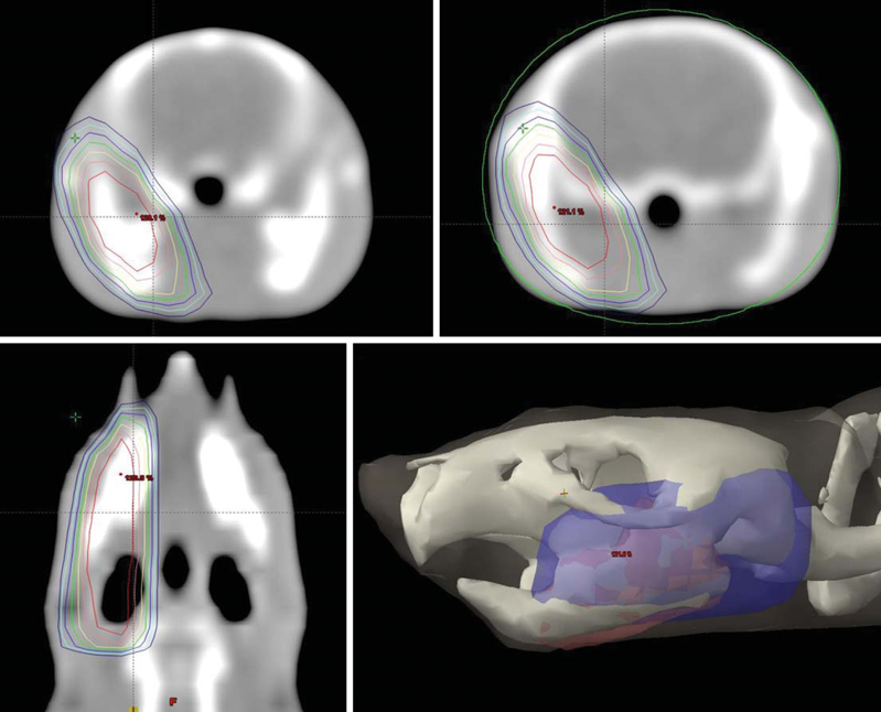
Fig. 1 Target volume of the right mandible including the ramus and condyle in CT scans are set prior to high-dose irradiation. 100% isodoses are marked in yellow. Pre-irradiated planning enables setting a one-sided application of radiation and minimizing the change in adjacent tissues, especially the brain.
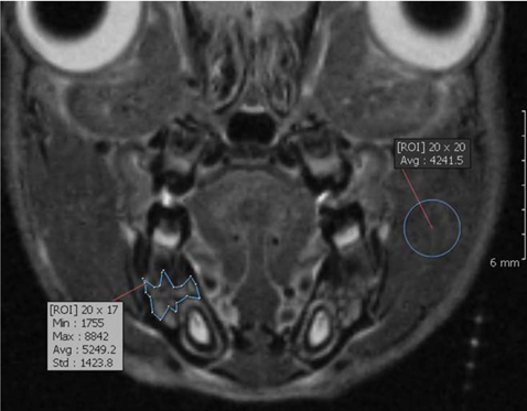
Fig. 2 Coronal fat-suppressed T2-weighted MR image. In order to locate the same plane every week, the axial (pulp floor of molar teeth), sagittal (midline of face), and coronal (first molar including two roots) planes were adjusted. The location of the region of interest (ROI) was used to measure the signal intensity in the mandibular first molar area. The ROIs included the alveolar bone marrow between the first molar and the incisor, and the adjacent muscles.
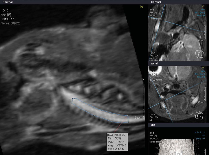
Fig. 3 The ROI of the incisor pulp tissue below the molar teeth on the modified sagittal section is obtained along the long axis of the incisor.
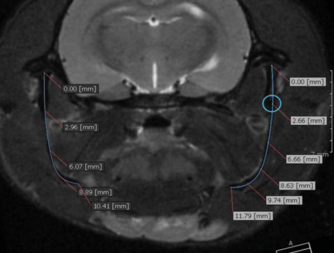
Fig. 4 Coronal fat-suppressed T2-weighted MR image. The image shows an example of a mandibular ramus height (condyle to mandibular angle) measurement using the Tapeline tool in the OnDemand3D program.
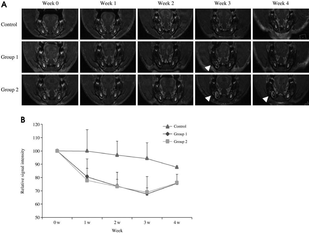
Fig. 5 A. The MR images show the changes during the 4 weeks after irradiation. The SI of the irradiated tissues around the right incisor pulp has increased (arrowhead). The changes in the MR images in Group 2 are prominent. B. The diagram shows the SI of the bone marrow versus the time curve. In the control group, the SI of the bone marrow has decreased. The SI of the irradiated right bone marrow has also decreased. The irradiated groups show a lower level of SI than that of the control group. These differences appear from the first week. However, a small difference can be seen between the Groups 1 and 2.
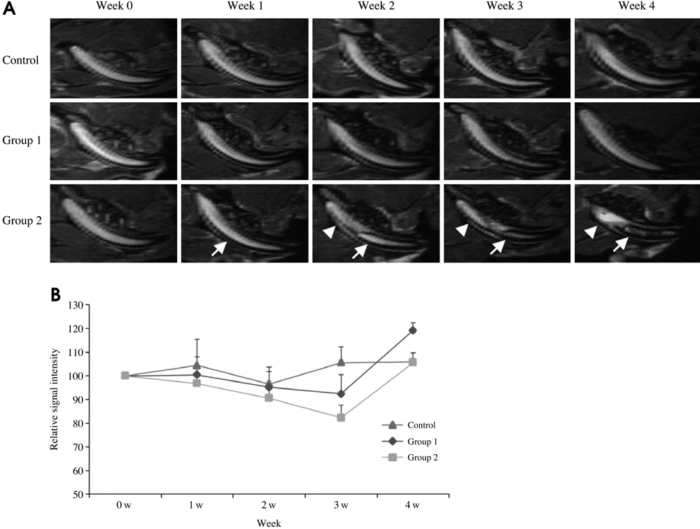
Fig. 6 A. The MR images show the changes during the 4 weeks after irradiation. A modified sagittal view is obtained along the long axis of the incisor. The size and SI of pulp of incisor have decreased (arrow) in Group 2. However, the SI of the MR images in the Group 2 is high at the apex of the incisor from week 2 after irradiation (arrowhead). B. The diagram shows the SI of the pulp tissue versus the time curve. The SI of the irradiated pulp tissue has decreased until week 3 after irradiation. There are differences among the groups at week 3.
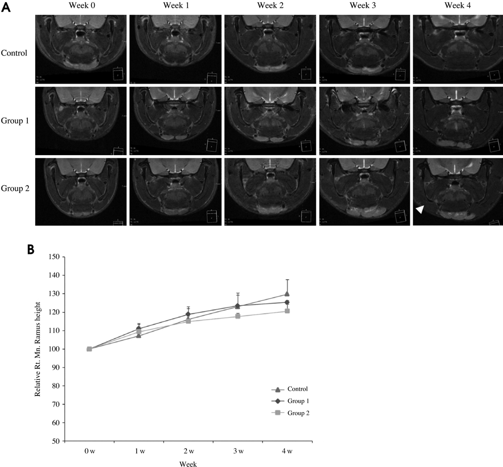
Fig. 7 A. The MR images show the changes during the 4 weeks after irradiation. There is a mandibular asymmetry due to the retardation of the growth on the right side (arrowhead). B. The diagram shows the mandibular ramus height versus the time curve. The height of the irradiated ramus is shorter than the contralateral and control side.
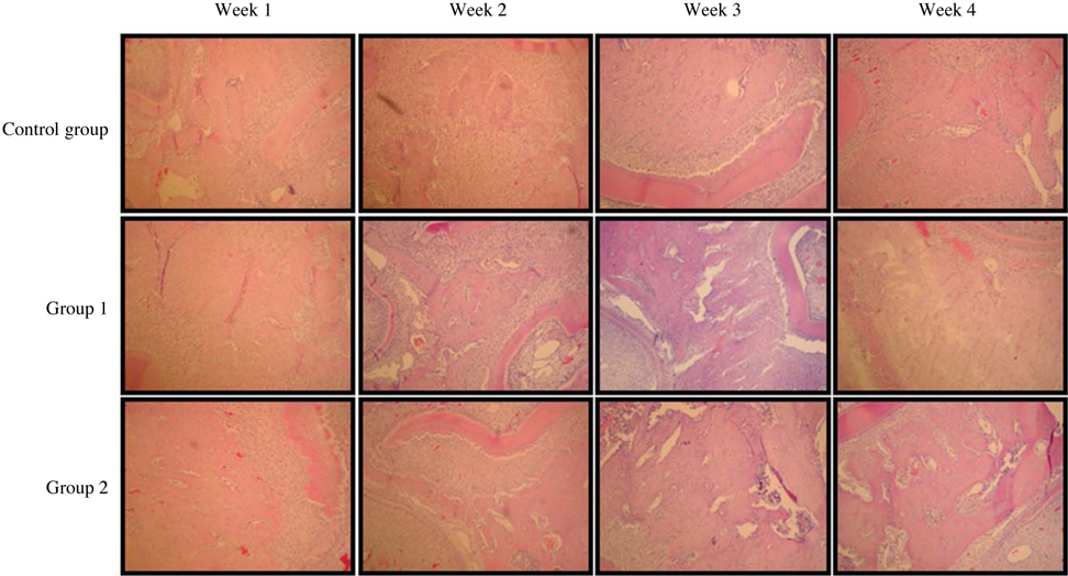
Fig. 8 Histopathologic examination of the alveolar bone below molar tooth (H&E stain, 100×). At the irradiated sites, the histopathologic changes show increasing trabecular bone formation and bone sclerosis in the irradiated groups, prominent in the molar region in Group 2.
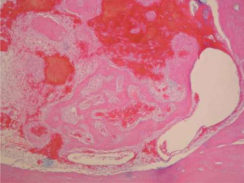
Fig. 9 Histopathologic changes show the atrophic incisor pulp tissue due to irradiation. The enlarged vessels and hemorrhagic changes are visible. The formation of dental hard tissue is inhibited, but it shows new bone formation (Group 2 at week 4 after irradiation. H&E stain, 100×).
Reference
-
1. Tofilon PJ, Fike JR. The radioresponse of the central nervous system: a dynamic process. Radiat Res. 2000; 153:357–370.
Article2. White SC, Pharoah MJ. Oral radiology; principles and interpretation. 5th ed. St. Louis: Mosby-Year Book;2004. p. 25–44.3. Matsumura S, Jikko A, Hiranuma H, Deguchi A, Fuchihata H. Effect of X-ray irradiation on proliferation and differentiation of osteoblast. Calcif Tissue Int. 1996; 59:307–308.
Article4. Sugimoto M, Takahashi S, Toguchida J, Kotoura Y, Shibamoto Y, Yamamuro T. Changes in bone after high-dose irradiation. Biomechanics and histomorphology. J Bone Joint Surg Br. 1991; 73:492–497.
Article5. Moore GS. Pediatric musculoskeletal imaging. In : Stark DS, Bradley WG, editors. Magnetic resonance imaging. 2nd ed. St. Louis: Mosby-Year Book;1992. p. 2223–2274.6. Niehoff P, Springer IN, Açil Y, Lange A, Marget M, Roldán JC, et al. HDR brachytherapy irradiation of the jaw - as a new experimental model of radiogenic bone damage. J Craniomaxillofac Surg. 2008; 36:203–209.
Article7. Vier-Pelisser FV, Figueiredo MA, Cherubini K, Braga Filho A, Figueiredo JA. The effect of head-fractioned teletherapy on pulp tissue. Int Endod J. 2007; 40:859–865.
Article8. Furstman LL. Effect of X irradiation on the mandibular condyle. J Dent Res. 1970; 49:419–427.
Article9. Tamplen M, Trapp K, Nishimura I, Armin B, Steinberg M, Beumer J, et al. Standardized analysis of mandibular osteoradionecrosis in a rat model. Otolaryngol Head Neck Surg. 2011; 145:404–410.
Article10. Bachmann G, Rössler R, Klett R, Rau WS, Bauer R. The role of magnetic resonance imaging and scintigraphy in the diagnosis of pathologic changes of the mandible after radiation therapy. Int J Oral Maxillofac Surg. 1996; 25:189–195.
Article11. Onu M, Savu M, Lungu-Solomonescu C, Harabagiu I, Pop T. Early MR changes in vertebral bone marrow for patients following radiotherapy. Eur Radiol. 2001; 11:1463–1469.
Article12. Stevens SK, Moore SG, Kaplan ID. Early and late bone-marrow changes after irradiation: MR evaluation. AJR Am J Roentgenol. 1990; 154:745–750.
Article13. Blomlie V, Rofstad EK, Skjønsberg A, Tverå K, Lien HH. Female pelvic bone marrow: serial MR imaging before, during, and after radiation therapy. Radiology. 1995; 194:537–543.
Article14. Store G, Larheim TA. Mandibular osteoradionecrosis: a comparison of computed tomography with panoramic radiography. Dentomaxillofac Radiol. 1999; 28:295–300.
Article15. Kaplan PA, Dussault RG. Magnetic resonance imaging of the bone marrow. In : Higgins CB, Hricak H, Helms CA, editors. Magnetic resonance imaging of the body. 3rd ed. Philadelphia: Lippincott-Raven;1997. p. 101–126.16. Vogler JB 3rd, Murphy WA. Bone marrow imaging. Radiology. 1988; 168:679–693.
Article17. Kaneda T, Minami M, Ozawa K, Akimoto Y, Okada H, Yamamoto H, et al. Magnetic resonance appearance of bone marrow in the mandible at different ages. Oral Surg Oral Med Oral Pathol Oral Radiol Endod. 1996; 82:229–233.
Article18. Unger E, Moldofsky P, Gatenby R, Hartz W, Broder G. Diagnosis of osteomyelitis by MR imaging. AJR Am J Roentgenol. 1988; 150:605–610.
Article19. Kaneda T, Minami M, Ozawa K, Akimoto Y, Utsunomiya T, Yamamoto H, et al. Magnetic resonance imaging of osteomyelitis in the mandible. Comparative study with other radiologic modalities. Oral Surg Oral Med Oral Pathol Oral Radiol Endod. 1995; 79:634–640.20. Weber-Donat G, Amabile JC, Lahutte-Auboin M, Potet J, Baccialone J, Bey E, et al. MRI assessment of local acute radiation syndrome. Eur Radiol. 2012; 22:2814–2821.
Article21. Sugimura H, Kisanuki A, Tamura S, Kihara Y, Watanabe K, Sumiyoshi A. Magnetic resonance imaging of bone marrow changes after irradiation. Invest Radiol. 1994; 29:35–41.
Article22. Tang JS, Gold RH, Bassett LW, Seeger LL. Musculoskeletal infection of the extremities: evaluation with MR imaging. Radiology. 1988; 166:205–209.
Article23. Nolte-Ernsting CC, Adam G, Bühne M, Prescher A, Günther RW. MRI of degenerative bone marrow lesions in experimental osteoarthritis of canine knee joints. Skeletal Radiol. 1996; 25:413–420.
Article24. Springer IN, Niehoff P, Açil Y, Marget M, Lange A, Warnke PH, et al. BMP-2 and bFGF in an irradiated bone model. J Craniomaxillofac Surg. 2008; 36:210–217.
Article25. Schultze-Mosgau S, Lehner B, Rödel F, Wehrhan F, Amann K, Kopp J, et al. Expression of bone morphogenic protein 2/4, transforming growth factor-beta1, and bone matrix protein expression in healing area between vascular tibia grafts and irradiated bone-experimental model of osteonecrosis. Int J Radiat Oncol Biol Phys. 2005; 61:1189–1196.26. Cohen M, Nishimura I, Tamplen M, Hokugo A, Beumer J, Steinberg ML, et al. Animal model of radiogenic bone damage to study mandibular osteoradionecrosis. Am J Otolaryngol. 2011; 32:291–300.
Article27. Little JB. Cellular, molecular, and carcinogenic effects of radiation. Hematol Oncol Clin North Am. 1993; 7:337–352.
Article
- Full Text Links
-
- Actions
-
Cited
- CITED
-
- Close
- Share
-
- Similar articles
-
- MR Imaging of the Bone Marrow
- Effect of irradiation on the temporomandibular joint in streptozotocin-induced diabetic rat
- Gelatinous Transformation of Bone Marrow Mimicking Malignant Marrow-Replacing Lesion on Magnetic Resonance Imaging in a Patient without Underlying Devastating Disease
- A Study on the Dose Distribution for Total Body Irradiation using Co-60 Teletherapy Unit
- Parotid mandibular bone defect: A case report emphasizing imaging features in plain radiographs and magnetic resonance imaging
