Solitary peripheral osteomas of the jaws
- Affiliations
-
- 1Department of Clinical and Preventive Dentistry, Oral Pathology Unit, Federal University of Pernambuco, Recife, Pernambuco, Brazil.
- 2Department of Clinical and Preventive Dentistry, Oral Medicine Unit, Federal University of Pernambuco, Recife, Pernambuco, Brazil. danyel.perez@ufpe.br
- 3Department of Oral and Maxillofacial Surgery, Hospital Geral de Areas, Recife, Pernambuco, Brazil.
- KMID: 1974416
- DOI: http://doi.org/10.5624/isd.2012.42.2.99
Abstract
- Osteoma is a benign osteogenic tumor composed of cancellous or compact bone, classified as peripheral, central, or extraskeletal. Peripheral osteomas are uncommon. Excluding the maxillary sinuses, the maxilla is a rare site for osteomas. The purpose of this report was to describe clinicopathological and radiological features of two peripheral osteomas occurring in the jaws, one located in the mandible and another in the edentulous maxillary alveolar ridge. The tumors were asymptomatic and were fully excised without any complications or recurrence. The lesions were submitted to histopathological analysis and diagnosed as peripheral osteoma, compact type.
Figure
-
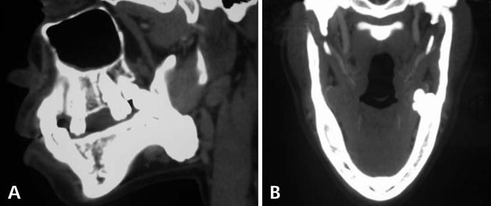
Fig. 1 A. Computed tomography (CT) scan reveals a well-circumscribed, bone-like, hyperdense image located on the left mandibular angle. B. CT image shows a lesion with a lobulated surface.
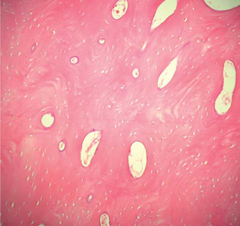
Fig. 2 Histopathological examination shows compact and mature bone, with scarce lacunae and marrow spaces filled by a connective tissue (H&E stain, ×100).
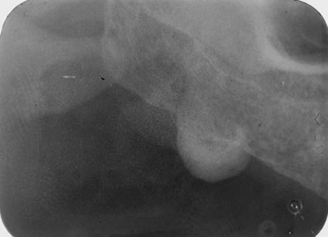
Fig. 3 Periapical radiograph shows a well delimited, ovoid, and radiopaque lesion observed on the edentulous alveolar ridge.
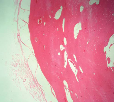
Fig. 4 Histopathological examination shows maxillary osteoma, compact type, circumscribed by a fibrous capsule around the lesion (H&E stain, ×100).
Cited by 1 articles
-
Giant complex odontoma in the posterior mandible: A case report and literature review
Jong Chan Park, Ji Ho Yang, Sung Youn Jo, Bong Chul Kim, Jun Lee, Wan Lee
Imaging Sci Dent. 2018;48(4):289-293. doi: 10.5624/isd.2018.48.4.289.
Reference
-
1. Sayan NB, Uçok C, Karasu HA, Günhan O. Peripheral osteoma of the oral and maxillofacial region: a study of 35 new cases. J Oral Maxillofac Surg. 2002. 60:1299–1301.
Article2. Aghabeigi B, Evans AW, Crean SJ, Hopper C. Simultaneous repair of an orbital floor fracture and removal of an ethmoid osteoma: case report and review of the literature. Int J Oral Maxillofac Surg. 2003. 32:94–96.
Article3. Durighetto AF Jr, de Moraes Ramos FM, Rocha MA, da Cruz Perez DE. Peripheral osteoma of the maxilla: report of a case. Dentomaxillofac Radiol. 2007. 36:308–310.
Article4. Longo F, Califano L, De Maria G, Ciccarelli R. Solitary osteoma of the mandibular ramus: report of a case. J Oral Maxillofac Surg. 2001. 59:698–700.
Article5. Kaplan I, Calderon S, Buchner A. Peripheral osteoma of the mandible: a study of 10 new cases and analysis of the literature. J Oral Maxillofac Surg. 1994. 52:467–470.
Article6. Bodner L, Gatot A, Sion-Vardy N, Fliss DM. Peripheral osteoma of the mandibular ascending ramus. J Oral Maxillofac Surg. 1998. 56:1446–1449.
Article7. Nah KS. Osteomas of the craniofacial region. Imaging Sci Dent. 2011. 41:107–113.
Article8. Seward MH. An osteoma of the maxilla. Br Dent J. 1965. 118:27–30.9. Rajayogeswaran V, Eveson JW. Endosteal (central) osteoma of the maxilla. Br Dent J. 1981. 150:162–163.
Article10. Kaplan I, Nicolaou Z, Hatuel D, Calderon S. Solitary central osteoma of the jaws: a diagnostic dilemma. Oral Surg Oral Med Oral Pathol Oral Radiol Endod. 2008. 106:e22–e29.
Article11. Dalambiras S, Boutsioukis C, Tilaveridis I. Peripheral osteoma of the maxilla: report of an unusual case. Oral Surg Oral Med Oral Pathol Oral Radiol Endod. 2005. 100:e19–e24.
Article12. Woldenberg Y, Nash M, Bodner L. Peripheral osteoma of the maxillofacial region. Diagnosis and management: a study of 14 cases. Med Oral Patol Oral Cir Bucal. 2005. 10:E139–E142.13. Iatrou IA, Leventis MD, Dais PE, Tosios KI. Peripheral osteoma of the maxillary alveolar process. J Craniofac Surg. 2007. 18:1169–1173.
Article14. Chaudhry J, Rawal SY, Anderson KM, Rawal YB. Cancellous osteoma of the maxillary tuberosity: case report. Gen Dent. 2009. 57:427–429.15. Wong RC, Peck RH. Enlargement of the right maxilla - report of an unusual peripheral osteoma. Ann Acad Med Singapore. 2010. 39:576–577.16. de Santana Santos T, Frota R, Martins-Filho PR, Melo AR, de Souza Andrade ES, de Oliveira e Silva ED, et al. Central osteoma of the maxilla with involvement of paranasal sinus. J Craniofac Surg. 2011. 22:589–591.17. Sah K, Kale A, Seema H, Kotrashetti V, Pramod BJ. Peripheral osteoma of the maxilla: a rare case report. Contemp Clin Dent. 2011. 2:49–52.
Article18. Cutilli BJ, Quinn PD. Traumatically induced peripheral osteoma. Report of a case. Oral Surg Oral Med Oral Pathol. 1992. 73:667–669.19. Kashima K, Rahman OI, Sakoda S, Shiba R. Unusual peripheral osteoma of the mandible: report of 2 cases. J Oral Maxillofac Surg. 2000. 58:911–913.
Article20. Neville B, Damm DD, Allen CM, Bouquot J. Oral and maxillofacial pathology. 2009. 3rd ed. St. Louis: Saunders;19–22.21. Verweij KE, Engelkens HJ, Bertheux CA, Dees A. Multiple lesions in upper jaw. Multiple buccal exostoses. Neth J Med. 2011. 69:347–350.22. Johann AC, de Freitas JB, de Aguiar MC, de Araújo NS, Mesquita RA. Peripheral osteoma of the mandible: case report and review of the literature. J Craniomaxillofac Surg. 2005. 33:276–281.
Article23. Payne M, Anderson JA, Cook J. Gardner's syndrome - a case report. Br Dent J. 2002. 193:383–384.
Article
