Imaging Sci Dent.
2011 Mar;41(1):17-21. 10.5624/isd.2011.41.1.17.
External root resorption after orthodontic treatment: a study of contributing factors
- Affiliations
-
- 1Department of Oral and Maxillofacial Radiology, School of Dentistry, Pusan National University, Pusan, Korea. bhjo@pusan.ac.kr
- KMID: 1974395
- DOI: http://doi.org/10.5624/isd.2011.41.1.17
Abstract
- PURPOSE
The purpose of this study was to examine the patient- and treatment-related etiologic factors of external root resorption.
MATERIALS AND METHODS
This study consisted of 163 patients who had completed orthodontic treatments and taken the pre- and post-treatment panoramic and lateral cephalometric radiographs. The length of tooth was measured from the tooth apex to the incisal edge or cusp tip on the panoramic radiograph. Overbite and overjet were measured from the pre- and post-treatment lateral cephalometric radiographs. The root resorption of each tooth and the factors of malocclusion were analyzed with an analysis of variance. A paired t test was performed to compare the mean amount of root resorption between male and female, between extraction and non-extraction cases, and between surgery and non-surgery groups. Correlation coefficients were measured to assess the relationship between the amount of root resorption and the age in which the orthodontic treatment started, the degree of changes in overbite and overjet, and the duration of treatment.
RESULTS
Maxillary central incisor was the most resorbed tooth, followed by the maxillary lateral incisor, the mandibular central incisor, and the mandibular lateral incisor. The history of tooth extraction was significantly associated with the root resorption. The duration of orthodontic treatment was positively correlated with the amount of root resorption.
CONCLUSION
These findings show that orthodontic treatment should be carefully performed in patients who need the treatment for a long period and with a pre-treatment extraction of teeth.
Keyword
MeSH Terms
Figure
-
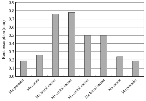
Fig. 1 The amount of root resorption by tooth type* (N=326†). *Significant differences for tooth types by ANOVA (P<0.01). †Data from the left and right sides were pooled to simplify presentation, so counts are of teeth, not person.
Cited by 1 articles
-
A posteriori registration and subtraction of periapical radiographs for the evaluation of external apical root resorption after orthodontic treatment
Eliane Maria Kreich, Ana Cláudia Chibinski, Ulisses Coelho, Letícia Stadler Wambier, Rosário de Arruda Moura Zedebski, Mari Eli Leonelli de Moraes, Luiz Cesar de Moraes
Imaging Sci Dent. 2016;46(1):17-24. doi: 10.5624/isd.2016.46.1.17.
Reference
-
1. Brudvik P, Rygh P. Non-clast cells start orthodontic root resorption in the periphery of hyalinized zones. Eur J Orthod. 1993. 15:467–480.
Article2. Brudvik P, Rygh P. Root resorption beneath the main hyalinized zone. Eur J Orthod. 1994. 16:249–263.
Article3. Kurol J, Owman-Moll P. Hyalinization and root resorption during early orthodontic tooth movement in adolescents. Angle Orthod. 1998. 68:161–165.4. Reitan K. Initial tissue behavior during apical root resorption. Angle Orthod. 1974. 44:68–82.5. Costopoulos G, Nanda R. An evaluation of root resorption incident to orthodontic intrusion. Am J Orthod Dentofacial Orthop. 1996. 109:543–548.
Article6. Segal GR, Schiffman PH, Tuncay OC. Meta analysis of the treatment-related factors of external apical root resorption. Orthod Craniofac Res. 2004. 7:71–78.
Article7. Brezniak N, Wasserstein A. Root resorption after orthodontic treatment: Part 1. Literature review. Am J Orthod Dentofacial Orthop. 1993. 103:62–66.
Article8. Harris EF. Root resorption during orthodontic therapy. Semin Orthod. 2000. 6:183–194.
Article9. Owman-Moll P, Kurol J, Lundgren D. Continuous versus interrupted continuous orthodontic force related to early tooth movement and root resorption. Angle Orthod. 1995. 65:395–402.10. McGuinness N, Wilson AN, Jones M, Middleton J, Robertson NR. Stresses induced by edgewise appliances in the periodontal ligament - a finite element study. Angle Orthod. 1992. 62:15–22.11. Kaimenyi JT, Ashley FP. Assessment of bone loss in periodontitis from panoramic radiographs. J Clin Periodontol. 1988. 15:170–174.
Article12. Sameshima GT, Sinclair PM. Predicting and preventing root resorption: Part I. Diagnostic factors. Am J Orthod Dentofacial Orthop. 2001. 119:505–510.
Article13. Linge BO, Linge L. Apical root resorption in upper anterior teeth. Eur J Orthod. 1983. 5:173–183.
Article14. Mirabella AD, Artun J. Prevalence and severity of apical root resorption of maxillary anterior teeth in adult orthodontic patients. Eur J Orthod. 1995. 17:93–99.
Article15. Harris EF, Baker WC. Loss of root length and crestal bone height before and during treatment in adolescent and adult orthodontic patients. Am J Orthod Dentofacial Orthop. 1990. 98:463–469.16. Baumrind S, Korn EL, Boyd RL. Apical root resorption in orthodontically treated adults. Am J Orthod Dentofacial Orthop. 1996. 110:311–320.
Article17. McNab S, Battistutta D, Taverne A, Symons AL. External apical root resorption following orthodontic treatment. Angle Orthod. 2000. 70:227–232.18. Parker RJ, Harris EF. Directions of orthodontic tooth movements associated with external apical root resorption of the maxillary central incisor. Am J Orthod Dentofacial Orthop. 1998. 114:677–683.
Article19. Sharpe W, Reed B, Subtelny JD, Polson A. Orthodontic relapse, apical root resorption, and crestal alveolar bone levels. Am J Orthod Dentofacial Orthop. 1987. 91:252–258.
Article20. Kaley J, Phillips C. Factors related to root resorption in edgewise practice. Angle Orthod. 1991. 61:125–132.21. Spurrier SW, Hall SH, Joondeph DR, Shapiro PA, Riedel RA. A comparison of apical root resorption during orthodontic treatment in endodontically treated and vital teeth. Am J Orthod Dentofacial Orthop. 1990. 97:130–134.
Article22. McFadden WM, Engstrom C, Engstrom H, Anholm JM. A study of the relationship between incisor intrusion and root shortening. Am J Orthod Dentofacial Orthop. 1989. 96:390–396.
Article23. Harris EF, Kineret SE, Tolley EA. A heritable component for external apical root resorption in patients treated orthodontically. Am J Orthod Dentofacial Orthop. 1997. 111:301–309.
Article24. Linge L, Linge BO. Patient characteristics and treatment variables associated with apical root resorption during orthodontic treatment. Am J Orthod Dentofacial Orthop. 1991. 99:35–43.
Article25. Sameshima GT, Sinclair PM. Predicting and preventing root resorption: Part II. Treatment factors. Am J Orthod Dentofacial Orthop. 2001. 119:511–515.
Article26. Levander E, Malmgren O. Evaluation of the risk of root resorption during orthodontic treatment: a study of upper incisors. Eur J Orthod. 1988. 10:30–38.
Article27. Kurol J, Owman-Moll P, Lundgren D. Time-related root resorption after application of a controlled continuous orthodontic force. Am J Orthod Dentofacial Orthop. 1996. 110:303–310.28. Mirabella AD, Artun J. Risk factors for apical root resorption of maxillary anterior teeth in adult orthodontic patients. Am J Orthod Dentofacial Orthop. 1995. 108:48–55.
Article29. Janson GR, De Luca Canto G, Martins DR, Henriques JF, De Freitas MR. A radiographic comparison of apical root resorption after orthodontic treatment with 3 different fixed appliance techniques. Am J Orthod Dentofacial Orthop. 2000. 118:262–273.
Article30. Vlaskalic V, Boyd RL, Baumrind S. Etiology and sequelae of root resorption. Semin Orthod. 1998. 4:124–131.
Article31. Beck BW, Harris EF. Apical root resorption in orthodontically treated subjects: analysis of edgewise and light wire mechanics. Am J Orthod Dentofacial Orthop. 1994. 105:350–361.
Article32. Otis LL, Hong JS, Tuncay OC. Bone structure effect on root resorption. Orthod Craniofac Res. 2004. 7:165–177.
Article33. Harris EF, Butler ML. Patterns of incisor root resorption before and after orthodontic correction in cases with anterior open bites. Am J Orthod Dentofacial Orthop. 1992. 101:112–119.
Article
- Full Text Links
-
- Actions
-
Cited
- CITED
-
- Close
- Share
-
- Similar articles
-
- A study on the affecting factors on root resorption
- Comparison between anterior segmental osteotomy versus conventional orthodontic treatment in root resorption: a radiographic study using cone-beam computed tomography
- External apical root resorption in maxillary incisors in orthodontic patients: associated factors and radiographic evaluation
- The effet of types of orthodontic force on the root resorption and repair in rat molar
- Changes in the titer of tooth root antibodies accompanying root resorption associated with orthodontic tooth movement
