The Usefulness of Three-dimensional Computed Tomography as an Assessment of Periacetabular Osteolysis in Revision Total Hip Arthroplasty
- Affiliations
-
- 1Department of Orthopedic Surgery, Inha University School of Medicine, Incheon, Korea. moon@inha.ac.kr
- KMID: 1974386
- DOI: http://doi.org/10.5371/hp.2015.27.2.90
Abstract
- PURPOSE
This study was performed to determine the usefulness of three-dimensional computed tomography (3D-CT) in measuring periacetabular osteolysis by comparing the real volume of osteolysis in revision surgery.
MATERIALS AND METHODS
Twnety-three patients who had undergone revision surgery due to periacetabular osteolysis but not included septic osteolysis and implant loosening. The mean age of patients at the time of surgery was 55.2 years. And the mean time interval between the primary total hip arthroplasty and revision surgery was 13.3 years. We measured the polyethylene wear in plain radiographs using computer assisted vector wear analysis program, the volume of acetabular osteolytic lesions in high-resolution spiral CT scans using Rapidia 3D software version 2.8 algorithms before the revision surgery were performed. Intraoperative real osteolytic volume was calculated as the sum of the volumetric increments of the acetabular cup and impacted allo-cancellous bone volume.
RESULTS
Strong correlation was found between the volume of acetabular osteolytic lesions measured on 3D-CT and intraoperative real osteolytic volume which was calculated as the sum of the volumetric increments of the acetabular cup and impacted allo-cancellous bone volume.
CONCLUSION
3D-CT is considered a useful method for assessing and measuring the volume of periacetabular osteolysis before revision surgery.
Keyword
MeSH Terms
Figure
-
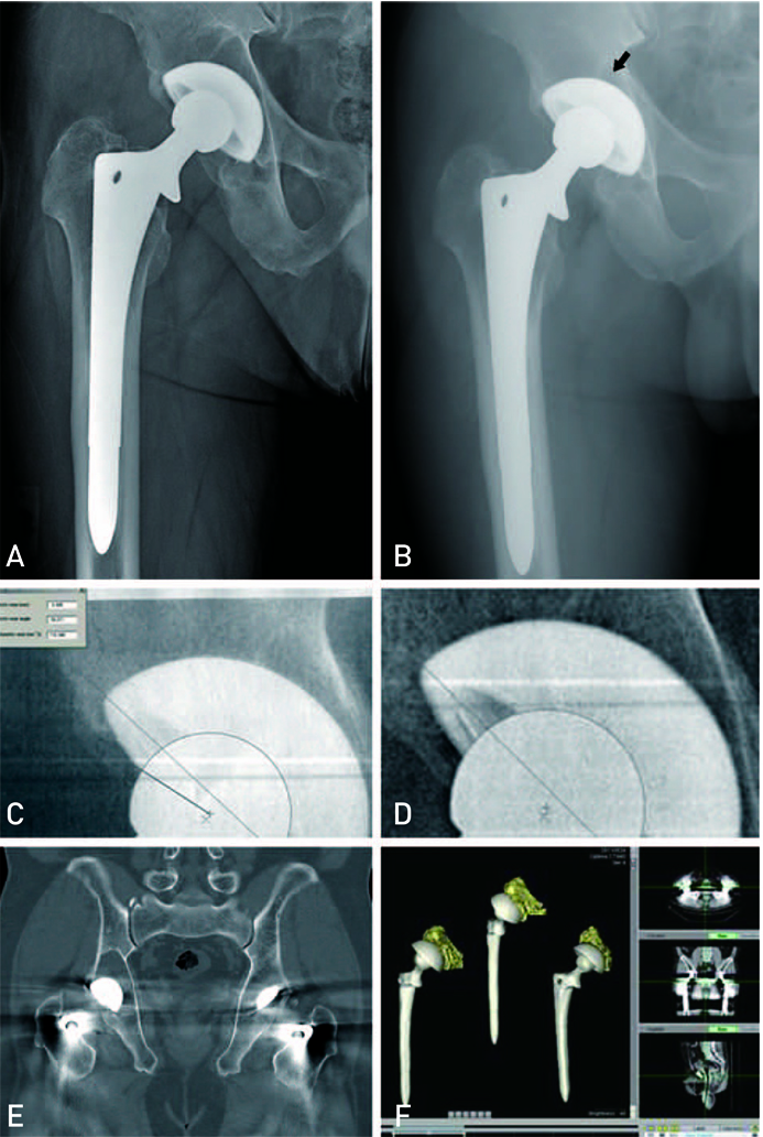
Fig. 1 This figure shows periacetabular osteolysis after primary total hip replacement arthroplasty in avascular necrosis of the right hip and measurement of osteolysis by computer assisted vector wear analysis program and three-dimentional (3D) computed tomography in a 56 years old man. (A) This immediate postoperative anteroposterior plain radiography shows good positioning of the prosthesis (May 2001). (B) After 10 years (May 2011), the plain radiography shows periacetabular osteolytic lesion (arrow). (C, D) The images were analyzed by applying a computer assisted vector wear analysis program (University of Chicago, hip analysis program 4.0). (E) We measured the volume of acetabular ostelytic lesions via high-resolution spiral computed tomography scans. (F) We reconstructed to a 3D ostelytic volume by using via Rapidia 3D software version 2.8.
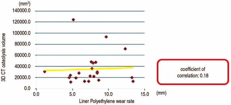
Fig. 2 A weak correlation was found between the mean linear wear rate of polyethylene liners measured on plain radiographs and osteolytic volumes measured on three-dimensional computed tomography (3D-CT) with no statistically significance (r2=0.18 , P=0.938).
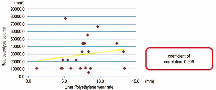
Fig. 3 The correlation between the mean linear and volumetric wear rate of polyethylene liners was weak with no statistically significance (r2=0.206, P=0.359).
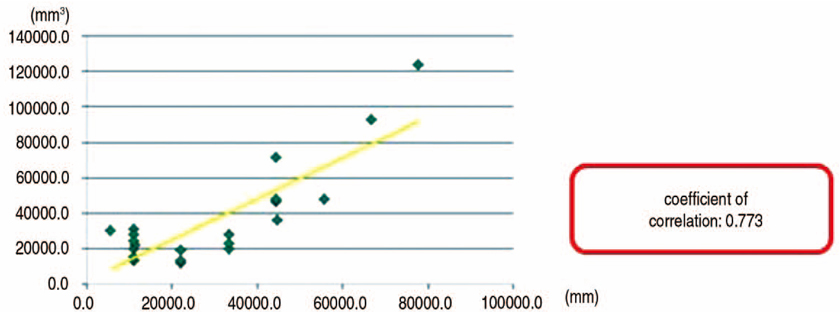
Fig. 4 A strong and significant relationship was found between osteolytic volume measured on the three-dimensional computed tomography and the sum of the volumetric increments of acetabular cups and impacted allo-cancellous bone volume with a statistical significance (r2=0.773, P<0.001).
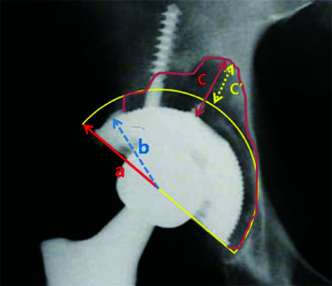
Fig. 5 The realationship between sum of the volumetric increments of acetabular cup and impacted allocancellous bone volume and osteolytic volume measured on three-dimensional computed tomography (3D-CT) (r=0.773, P<0.001). a: radius of acetabular wall, b: radius of acetabular cup, c: osteolytic volume measured on 3D-CT, c': grafted allo-cancellous bone volume.
Reference
-
1. Engh CA Jr, Culpepper WJ 2nd, Engh CA. Long-term results of use of the anatomic medullary locking prosthesis in total hip arthroplasty. J Bone Joint Surg Am. 1997; 79:177–184.
Article2. Harris WH. Wear and periprosthetic osteolysis: the problem. Clin Orthop Relat Res. 2001; 393:66–70.3. Maloney WJ, Galante JO, Anderson M, et al. Fixation, polyethylene wear, and pelvic osteolysis in primary total hip replacement. Clin Orthop Relat Res. 1999; 369:157–164.
Article4. Maloney WJ, Herzwurm P, Paprosky W, Rubash HE, Engh CA. Treatment of pelvic osteolysis associated with a stable acetabular component inserted without cement as part of a total hip replacement. J Bone Joint Surg Am. 1997; 79:1628–1634.
Article5. Xenos JS, Hopkinson WJ, Callaghan JJ, Heekin RD, Savory CG. Osteolysis around an uncemented cobalt chrome total hip arthroplasty. Clin Orthop Relat Res. 1995; 317:29–36.6. Carter DR, Hayes WC. The compressive behavior of bone as a two-phase porous structure. J Bone Joint Surg Am. 1977; 59:954–962.
Article7. Zicat B, Engh CA, Gokcen E. Patterns of osteolysis around total hip components inserted with and without cement. J Bone Joint Surg Am. 1995; 77:432–439.
Article8. Schmalzried TP, Guttmann D, Grecula M, Amstutz HC. The relationship between the design, position, and articular wear of acetabular components inserted without cement and the development of pelvic osteolysis. J Bone Joint Surg Am. 1994; 76:677–688.
Article9. Claus AM, Engh CA Jr, Sychterz CJ, Xenos JS, Orishimo KF, Engh CA Sr. Radiographic definition of pelvic osteolysis following total hip arthroplasty. J Bone Joint Surg Am. 2003; 85-A:1519–1526.
Article10. Kitamura N, Pappedemos PC, Duffy PR 3rd, et al. The value of anteroposterior pelvic radiographs for evaluating pelvic osteolysis. Clin Orthop Relat Res. 2006; 453:239–245.
Article11. Looney RJ, Boyd A, Totterman S, et al. Volumetric computerized tomography as a measurement of periprosthetic acetabular osteolysis and its correlation with wear. Arthritis Res. 2002; 4:59–63.12. Puri L, Wixson RL, Stern SH, Kohli J, Hendrix RW, Stulberg SD. Use of helical computed tomography for the assessment of acetabular osteolysis after total hip arthroplasty. J Bone Joint Surg Am. 2002; 84-A:609–614.
Article13. Stulberg SD, Wixson RL, Adams AD, Hendrix RW, Bernfield JB. Monitoring pelvic osteolysis following total hip replacement surgery: an algorithm for surveillance. J Bone Joint Surg Am. 2002; 84-A:Suppl 2. 116–122.14. Oparaugo PC, Clarke IC, Malchau H, Herberts P. Correlation of wear debris-induced osteolysis and revision with volumetric wear-rates of polyethylene: a survey of 8 reports in the literature. Acta Orthop Scand. 2001; 72:22–28.
Article15. Orishimo KF, Claus AM, Sychterz CJ, Engh CA. Relationship between polyethylene wear and osteolysis in hips with a second-generation porous-coated cementless cup after seven years of follow-up. J Bone Joint Surg Am. 2003; 85-A:1095–1099.16. Perez RE, Rodriguez JA, Deshmukh RG, Ranawat CS. Polyethylene wear and periprosthetic osteolysis in metal-backed acetabular components with cylindrical liners. J Arthroplasty. 1998; 13:1–7.
Article17. Martell JM, Berdia S. Determination of polyethylene wear in total hip replacements with use of digital radiographs. J Bone Joint Surg Am. 1997; 79:1635–1641.
Article18. Egawa H, Powers CC, Beykirch SE, Hopper RH Jr, Engh CA Jr, Engh CA. Can the volume of pelvic osteolysis be calculated without using computed tomography? Clin Orthop Relat Res. 2009; 467:181–187.
Article19. Howie DW, Neale SD, Stamenkov R, McGee MA, Taylor DJ, Findlay DM. Progression of acetabular periprosthetic osteolytic lesions measured with computed tomography. J Bone Joint Surg Am. 2007; 89:1818–1825.
Article20. Devane PA, Robinson EJ, Bourne RB, Rorabeck CH, Nayak NN, Horne JG. Measurement of polyethylene wear in acetabular components inserted with and without cement. A randomized trial. J Bone Joint Surg Am. 1997; 79:682–689.
Article21. Martell JM, Berkson E, Berger R, Jacobs J. Comparison of two and three-dimensional computerized polyethylene wear analysis after total hip arthroplasty. J Bone Joint Surg Am. 2003; 85-A:1111–1117.
Article22. McCalden RW, Naudie DD, Yuan X, Bourne RB. Radiographic methods for the assessment of polyethylene wear after total hip arthroplasty. J Bone Joint Surg Am. 2005; 87:2323–2334.
Article
- Full Text Links
-
- Actions
-
Cited
- CITED
-
- Close
- Share
-
- Similar articles
-
- A Comparison of Osteolysis in Bipolar Hemiarthroplasty and Total Hip Arthroplasty
- Revision Total Hip Arthroplasty of an Acetabular Cup with Acetabular Bone Defects
- Metal Debris Induced Osteolysis after Cementless Total Hip Arthroplasty: One case report
- Cementless Hip Arthroplasty in Patients with Avascular Necrosis of the Femoral Head: Long term results in AML
- Cementless Revision Total Hip Arthroplasty with Ceramic Articulation
