Maxillary Sinus Retention Cysts Protruding Into the Inferior Meatus
- Affiliations
-
- 1Department of Otorhinolaryngology-Head and Neck Surgery, Pusan National University Hospital, Busan, Korea. st-dragonhong@hanmail.net
- 2Biomedical Research Institute, Pusan National University Hospital, Busan, Korea.
- 3Department of Otorhinolaryngology-Head and Neck Surgery, Pusan National University Yangsan Hospital, Yangsan, Korea.
- KMID: 1973475
- DOI: http://doi.org/10.3342/ceo.2014.7.3.226
Abstract
- Although most of the maxillary sinus retention cysts are asymptomatic, a few of them increase in size and cause symptoms. However, they rarely erode bony walls nor protrude into the inferior meatus. I present 2 cases with maxillary sinus retention cysts protruding into the inferior meatus by making a large defect on the medial wall of the maxillary sinus.
Keyword
Figure
-
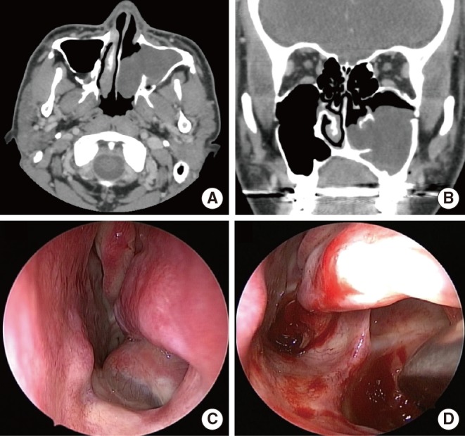
Fig. 1 Findings of computed tomogram and nasal endoscopy in the first patient. There is a non-enhancing mass occupying most of maxillary and protruding into the inferior meatus through the defect on the medial wall of maxillary sinus (MS) (A, B). He had osteotomy lines on the lateral walls of both MSs (B). Endoscopic examination revealed a cystic mass occupying the left inferior meatus (C). There remained a large defect with a clear margin on the medial wall of MS (D).
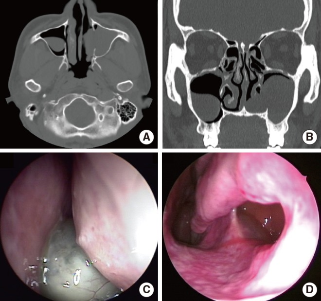
Fig. 2 Findings of computed tomogram and nasal endoscopy in the second patient. There is a non-enhancing mass occupying most of maxillary and protruding into the inferior meatus through the defect on the medial wall of maxillary sinus (MS) (A, B). He had a retention cyst confined to the right MS and a 4 mm-sized hole on the medial wall of this sinus (B). Endoscopic examination revealed a cystic mass occupying the left inferior meatus (C). There remained a large defect with a clear margin on the medial wall of MS (D).
Cited by 2 articles
-
A Case of Symptomatic Maxillary Retention Cyst
Hankyeol Kim, Eun Kyu Lee, Hyo Yeol Kim, Sang-Duck Hong, Hun-Jong Dhong, Seung-Kyu Chung
J Rhinol. 2018;25(1):59-62. doi: 10.18787/jr.2018.25.1.59.A Giant Maxillary Mucocele Presenting Left Cheek Swelling
Moon Seung Baeg, Hyeok Ro Kwon, Jin Soon Chang
J Rhinol. 2022;29(3):172-175. doi: 10.18787/jr.2022.00413.
Reference
-
1. Moon IJ, Kim SW, Han DH, Shin JM, Rhee CS, Lee CH, et al. Mucosal cysts in the paranasal sinuses: long-term follow-up and clinical implications. Am J Rhinol Allergy. 2011; Mar-Apr. 25(2):98–102. PMID: 21679512.
Article2. Składzien J, Litwin JA, Nowogrodzka-Zagorska M, Wierzchowski W. Morphological and clinical characteristics of antrochoanal polyps: comparison with chronic inflammation-associated polyps of the maxillary sinus. Auris Nasus Larynx. 2001; 4. 28(2):137–141. PMID: 11240321.
Article3. Moon IJ, Lee JE, Kim ST, Han DH, Rhee CS, Lee CH, et al. Characteristics and risk factors of mucosal cysts in the paranasal sinuses. Rhinology. 2011; 8. 49(3):309–314. PMID: 21858261.
Article4. Bósio JA, Tanaka O, Rovigatti E, de Gruner SK. The incidence of maxillary sinus retention cysts in orthodontic patients. World J Orthod. 2009; Summer. 10(2):e7–e8. PMID: 19582248.
- Full Text Links
-
- Actions
-
Cited
- CITED
-
- Close
- Share
-
- Similar articles
-
- Clinical Characteristics According to the Radiological Classifications of Maxillary Sinus Fungus Ball
- Treatment Strategy for the Retention Cyst of the Maxillary Sinus
- Sinus lifts in the presence of pseudoantral and mucous retention cysts
- Inferior Meatal Fenestration Operation of the Postoperative Maxillary Cysts
- Correlation of Middle Meatus, Ethmoid Sinus and Maxillary Sinus Microbiology in Patients with Chronic Sinusitis
