J Korean Neurosurg Soc.
2015 Apr;57(4):276-282. 10.3340/jkns.2015.57.4.276.
Quantification of Pediatric Cervical Spine Growth at the Cranio-Vertebral Junction
- Affiliations
-
- 1Department of Neurosurgery, Incheon St. Mary's Hospital, The Catholic University of Korea, Incheon, Korea. kjtns@olmh.cuk.ac.kr
- 2Department of Neurosurgery, St. Vincent's Hospital, The Catholic University of Korea, Suwon, Korea.
- KMID: 1956426
- DOI: http://doi.org/10.3340/jkns.2015.57.4.276
Abstract
OBJECTIVE
The purpose of this study was to investigate morphological change at the craniovertebral junction (CVJ) region using computed tomography.
METHODS
A total of 238 patients were included in this study, and mean age was 47.8+/-21.3 months. Spinal canal diameter, Power's ratio, McRae line, antero-posterior C1 ring height, atlantoaxial joint space, C2 growth, epidural space from the dens (M-PB-C2) and longitudinal distance (basion to C2 lower margin, B-C2) were measured. The mean value of each parameter was assessed for individual age groups. The cohorts were then divided into three larger age groups : infancy (I) (< or =2 years), very early (VE) childhood (2-5 years) and early (E) childhood (5> or = years).
RESULTS
Spinal canal diameter increased with age; however, this value did not increase with statistical significance after VE age. A significant age-related difference was found for all C2 body and odontoid parameters (p<0.05). Mean McRae line was 8.5, 8, and 7.5 mm in the I, VE, and E groups, respectively. The M-PB-C2 line showed up-and-down dynamic change during early pediatric periods.
CONCLUSION
Expansion of the spinal canal was restricted to the very early childhood period (less than 5 years) in the CVJ region; however, the C2 body and odontoid process increased continuously with age. The above results induced a dynamic change in the M-PB-C2 line. Although C2 longitudinal growth continued with age, the McRae line showed relatively little change.
Figure
-
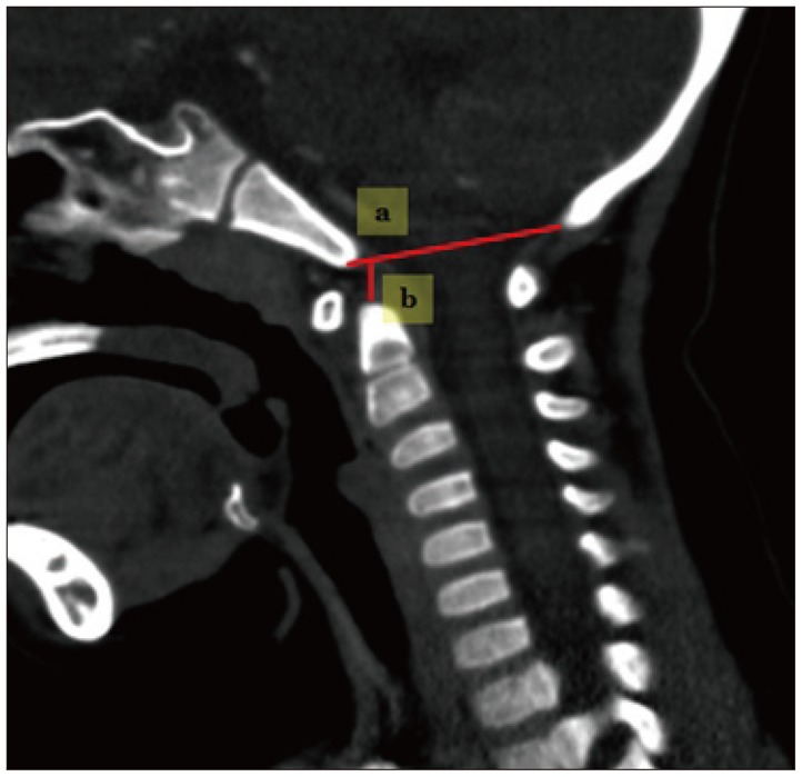
Fig. 1 Techniques for measuring the occipital area. a : occiput spinal canal antero-posterior diameter, b : McRae line.
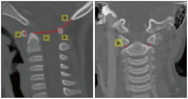
Fig. 2 Techniques for measuring in C1. a : C1 anterior arch height, b : C1 posterior arch height, c : C1 spinal canal antero-posterior diameter (C1-AP), d : occiput-C1 posterior arch distance (O-C1 interval), e : atlantoaxial (AA) joint space.

Fig. 3 Measuring techniques in C2. a : odontoid process height, b : odontoid process antero-posterior diameter, c : C2 body AP diameter, d : C2 body height, e : B-C2 distance, f : modified-(perpendicular basion-C2 line).
Reference
-
1. Chamoun RB, Whitehead WE, Curry DJ, Luerssen TG, Jea A. Computed tomography morphometric analysis for C-1 lateral mass screw placement in children. Clinical article. J Neurosurg Pediatr. 2009; 3:20–23. PMID: 19119899.
Article2. Cokluk C, Aydin K, Rakunt C, Iyigun O, Onder A. The borders of the odontoid process of C2 in adults and in children including the estimation of odontoid/body ratio. Eur Spine J. 2006; 15:278–282. PMID: 15968528.
Article3. Doherty BJ, Heggeness MH. Quantitative anatomy of the second cervical vertebra. Spine (Phila Pa 1976). 1995; 20:513–517. PMID: 7604318.
Article4. Fesmire FM, Luten RC. The pediatric cervical spine : developmental anatomy and clinical aspects. J Emerg Med. 1989; 7:133–142. PMID: 2661668.
Article5. Geck MJ, Truumees E, Hawthorne D, Singh D, Stokes JK, Flynn A. Feasibility of rigid upper cervical instrumentation in children : tomographic analysis of children aged 2-6. J Spinal Disord Tech. 2014; 27:E110–E117. PMID: 23563351.6. Grabb PA, Mapstone TB, Oakes WJ. Ventral brain stem compression in pediatric and young adult patients with Chiari I malformations. Neurosurgery. 1999; 44:520–527. discussion 527-528. PMID: 10069589.
Article7. Heller JG, Alson MD, Schaffler MB, Garfin SR. Quantitative internal dens morphology. Spine (Phila Pa 1976). 1992; 17:861–866. PMID: 1523487.
Article8. Lin SL, Xia DD, Chen W, Li Y, Shen ZH, Wang XY, et al. Computed tomographic morphometric analysis of the pediatric occipital condyle for occipital condyle screw placement. Spine (Phila Pa 1976). 2014; 39:E147–E152. PMID: 24173015.
Article9. Mcrae DL, Barnum AS. Occipitalization of the atlas. Am J Roentgenol Radium Ther Nucl Med. 1953; 70:23–46.10. Nucci RC, Seigal S, Merola AA, Gorup J, Mroczek KJ, Dryer J, et al. Computed tomographic evaluation of the normal adult odontoid. Implications for internal fixation. Spine (Phila Pa 1976). 1995; 20:264–270. PMID: 7732463.11. Piatt JH Jr, Grissom LE. Developmental anatomy of the atlas and axis in childhood by computed tomography. J Neurosurg Pediatr. 2011; 8:235–243. PMID: 21882912.
Article12. Powers B, Miller MD, Kramer RS, Martinez S, Gehweiler JA Jr. Traumatic anterior atlanto-occipital dislocation. Neurosurgery. 1979; 4:12–17. PMID: 450210.
Article13. Radcliff KE, Ben-Galim P, Dreiangel N, Martin SB, Reitman CA, Lin JN, et al. Comprehensive computed tomography assessment of the upper cervical anatomy : what is normal? Spine J. 2010; 10:219–229. PMID: 20207332.
Article14. Rao RD, Tang S, Lim C, Yoganandan N. Developmental morphology and ossification patterns of the C1 vertebra. J Bone Joint Surg Am. 2013; 95:e1241–e1247. PMID: 24005208.
Article15. Rijken BF, Lequin MH, de Rooi JJ, van Veelen ML, Mathijssen IM. Foramen magnum size and involvement of its intraoccipital synchondroses in Crouzon syndrome. Plast Reconstr Surg. 2013; 132:993e–1000e.
Article16. Sawada H, Akiguchi I, Fukuyama H, Kameyama M, Koyama T. Marked canal stenosis at the level of the atlas. Neuroradiology. 1983; 31:346–348. PMID: 2797428.
Article17. Schaffler MB, Alson MD, Heller JG, Garfin SR. Morphology of the dens. A quantitative study. Spine (Phila Pa 1976). 1992; 17:738–743. PMID: 1502635.
Article18. Tokiyoshi K, Nakagawa H, Kadota T. Spinal canal stenosis at the level of the atlas : case report. Surg Neurol. 1994; 41:238–240. PMID: 8146741.19. Tubbs RS, Wellons JC 3rd, Blount JP, Grabb PA, Oakes WJ. Inclination of the odontoid process in the pediatric Chiari I malformation. J Neurosurg. 2003; 98(1 Suppl):43–49. PMID: 12546387.
Article20. Yoganandan N, Pintar FA, Lew SM, Rao RD, Rangarajan N. Quantitative analyses of pediatric cervical spine ossification patterns using computed tomography. Ann Adv Automot Med. 2011; 55:159–168. PMID: 22105393.
- Full Text Links
-
- Actions
-
Cited
- CITED
-
- Close
- Share
-
- Similar articles
-
- Quantification of Pediatric Cervical Growth: Anatomical Changes in the Sub-Axial Spine
- Sagittal Fracture of Cervical Spine: Case Report
- Vertebral Artery Injury Following Blunt Trauma to the Cervical Spine Case Report and Literature Review
- Vertebral Artery Injury during Anterior Cervical Spine Surgery: Report of Two Cases
- Maturation of cervical vertebrae and mandibular growth changes
