The Combination of Neuroendocrine Tumor and Mucinous Neoplasm of the Appendix: A Case Report
- Affiliations
-
- 1Department of Radiology, Pusan National University Hospital, Pusan National University School of Medicine, Busan, Korea. leenk77@hanmail.net
- 2Department of Pathology, Pusan National University Hospital, Pusan National University School of Medicine, Busan, Korea.
- 3Department of Surgery, Pusan National University Hospital, Pusan National University School of Medicine, Busan, Korea.
- KMID: 1941781
- DOI: http://doi.org/10.3348/jksr.2014.70.5.355
Abstract
- Primary neoplasm of the appendix is an uncommon pathology, representing 0.5-1% of all appendix specimens. Especially, simultaneous occurrence of two tumors of the appendix was rarely documented. We report a case of the concomitant neuroendocrine tumor and the mucinous neoplasm of the appendix on abdominal computed tomography, in a 62-year-old female who came for a check-up.
Figure
-
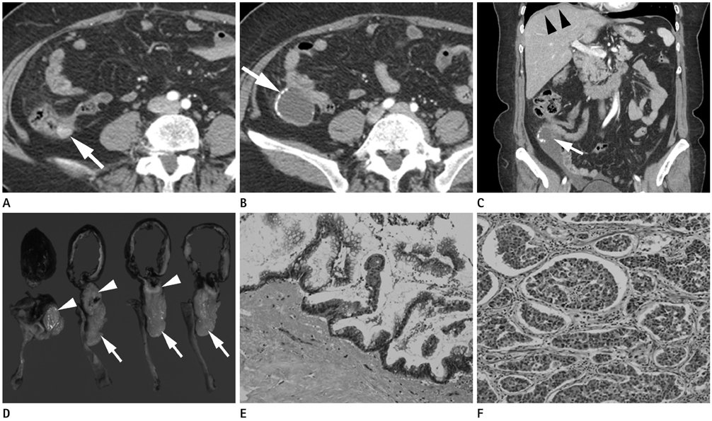
Fig. 1 A 62-year-old woman who presented with a neuroendocrine tumor and mucinous cystadenoma in the appendix. A. Axial contrast-enhanced CT shows a 1.1 cm sized well-defined enhancing mass (arrow) in the orifice of the appendix. B. Axial contrast-enhanced CT (caudal than A) shows a cystic mass (arrow) with peripheral calcification contiguous with base of cecum. C. Coronal-reformatted contrast-enhanced CT shows the rupture of the cystic mass (arrow) and periappendiceal fluid collection. Scalloping of the liver margin (arrowheads) is noted. D. Photograph of the cut gross specimens reveals cystic dilation of the appendix (arrows) and a subserosal mass (arrowheads) at the orifice of the appendix. E. Photomicrograph (original magnification, × 20 hematoxylin-eosin stain) of cystic dilation in the appendix shows replaced neoplastic mucinous epithelium with single layers, sometimes with papillary tufting. Nuclei are small and regular with low grade dysplasia. F. Photomicrograph (original magnification, × 20 hematoxylin-eosin stain) of the mass in the orifice of the appendix shows neoplastic neuroendocrine cells arranged in rounded solid nests. Tumor cells show moderate pleomorphism with occasionally seen hyperchromatic and large nuclei.
Reference
-
1. Pickhardt PJ, Levy AD, Rohrmann CA Jr, Kende AI. Primary neoplasms of the appendix: radiologic spectrum of disease with pathologic correlation. Radiographics. 2003; 23:645–662.2. Cho JB, Cho HJ, Shin OR, Kim KH, Ahn CH, Kim JS, et al. Clinical findings of appendiceal mucocele. J Korean Surg Soc. 2008; 74:429–435.3. Paik BL, Kim AR, Lee JH, Lee SB, Kim H, Park SJ, et al. The analysis of appendiceal mucocele with emphasis on the colonoscopic findings. Korean J Gastrointest Endosc. 2005; 30:243–248.4. Stocchi L, Wolff BG, Larson DR, Harrington JR. Surgical treatment of appendiceal mucocele. Arch Surg. 2003; 138:585–589. discussion 589-590.5. Brustmann H. Myxoglobulosis of the appendix associated with a proximal carcinoid and a pseudodiverticulum. Ann Diagn Pathol. 2006; 10:166–168.6. Alsaad KO, Serra S, Chetty R. Combined goblet cell carcinoid and mucinous cystadenoma of the vermiform appendix. World J Gastroenterol. 2009; 15:3431–3433.7. Rakovich G, Larue N. Mucocele of the appendix associated with a carcinoid of the terminal ileum. Can J Surg. 2007; 50:66–67.8. Carr NJ, Remotti H, Sobin LH. Dual carcinoid/epithelial neoplasia of the appendix. Histopathology. 1995; 27:557–562.9. Chang S, Choi D, Lee SJ, Lee WJ, Park MH, Kim SW, et al. Neuroendocrine neoplasms of the gastrointestinal tract: classification, pathologic basis, and imaging features. Radiographics. 2007; 27:1667–1679.10. Griniatsos J, Michail O. Appendiceal neuroendocrine tumors: recent insights and clinical implications. World J Gastrointest Oncol. 2010; 2:192–196.
- Full Text Links
-
- Actions
-
Cited
- CITED
-
- Close
- Share
-
- Similar articles
-
- Pancreatic Collision Tumor of Desmoid-Type Fibromatosis and Mucinous Cystic Neoplasm: A Case Report
- A Case of Pseudomyxoma Peritonei with Primary Borderline Mucinous Tumors of the Ovary and Appendix
- Ovarian Large Cell Neuroendocrine Carcinoma Associated with Endocervical-like Mucinous Borderline Tumor: A Case Report and Literature Review
- Gastrointestinal Stromal Tumor of the Appendix Mimicking a Mucinous Cystadenocarcinoma: A Case Report
- Incidental Carcinoid of Appendix in Borderline Mucinous Ovarian Tumor
