CT and MR Imaging Findings of Perianal Dermatofibrosarcoma Protuberans Mimicking Mucinous Adenocarcinoma Arising from Fistula in Ano: A Case Report
- Affiliations
-
- 1Department of Radiology, Uijeongbu St. Mary's Hospital, College of Medicine, The Catholic University of Korea, Uijeongbu, Korea. ymiku@catholic.ac.kr
- 2Department of Pathology, Uijeongbu St. Mary's Hospital, College of Medicine, The Catholic University of Korea, Uijeongbu, Korea.
- 3Department of Surgery, Uijeongbu St. Mary's Hospital, College of Medicine, The Catholic University of Korea, Uijeongbu, Korea.
- KMID: 1941772
- DOI: http://doi.org/10.3348/jksr.2014.70.6.435
Abstract
- Dermatofibrosarcoma protuberans (DFSP) is an uncommon soft tissue neoplasm which typically originates in the dermis and spreads into the subcutaneous layer and muscle. We report an extremely rare case of a perianal DFSP mimicking mucinous adenocarcinoma arising from anorectal fistula in a 59-year-old woman. In case of a perianal mass, which is connected by a stalk to the anus and shows heterogeneous enhancement without related regional lymphadenopathy, DFSP should be included in the differential diagnosis.
MeSH Terms
Figure
-
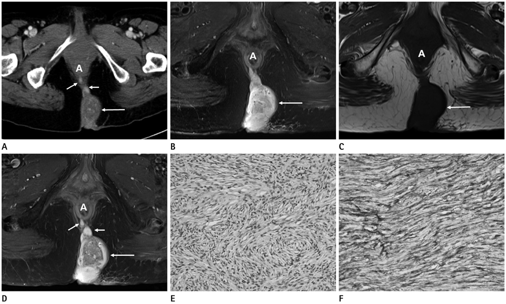
Fig. 1 A 59-year-old woman with perianal dermatofibrosarcoma protuberans (DFSP). A. Axial enhanced CT image shows a heterogenously enhancing mass (long arrow) in the medial aspect of the subcutaneous layer of the left gluteal region and with a communicating stalk (short arrows) between the mass and the anus, measuring 5.8 cm in size. B, C. The mass (arrows) shows bright signal intensity on fat-suppressed, T2-weighted imaging (B) and iso-signal intensity to muscle on T1-weighted imaging (C). D. Contrast-enhanced, fat-suppressed, T1-weighted MR images reveal heterogenous enhancement of the mass (long arrow) and the connecting stalk (short arrows). E. Photomicrograph shows interwoven fascicles of cells forming a storiform pattern (H&E, × 100). F. Immunohistochemical staining is strongly positive for CD34. These findings have been considered characteristic for the diagnosis of DFSP (CD34 DAB, × 400).
Reference
-
1. Llombart B, Serra-Guillén C, Monteagudo C, López Guerrero JA, Sanmartín O. Dermatofibrosarcoma protuberans: a comprehensive review and update on diagnosis and management. Semin Diagn Pathol. 2013; 30:13–28.2. Torreggiani WC, Al-Ismail K, Munk PL, Nicolaou S, O'Connell JX, Knowling MA. Dermatofibrosarcoma protuberans: MR imaging features. AJR Am J Roentgenol. 2002; 178:989–993.3. Mc Cormack O, Heaslip IS, Abdulrahim M, Murphy MN, Schranz M, O'Mahony JB. Perianal dermatofibrosarcoma protuberans: a case report. Cutis. 2011; 87:85–88.4. Kransdorf MJ, Meis-Kindblom JM. Dermatofibrosarcoma protuberans: radiologic appearance. AJR Am J Roentgenol. 1994; 163:391–394.5. Miyakawa E, Fujimoto H, Miyakawa K, Nemoto K, Kozawa K, Sugano I, et al. Dermatofibrosarcoma protuberans. CT findings with pathologic correlation in 6 cases. Acta Radiol. 1996; 37(3 Pt 1):362–365.6. Li X, Zhang W, Xiao L, Zeng Q, Wu X, Qin J, et al. Computed tomographic and pathological findings of dermatofibrosarcoma protuberans. J Comput Assist Tomogr. 2012; 36:462–468.7. Bergin P, Rezaei S, Lau Q, Coucher J. Dermatofibrosarcoma protuberans, magnetic resonance imaging and pathological correlation. Australas Radiol. 2007; 51 Spec No:B64–B66.8. Fujimoto H, Ikeda M, Shimofusa R, Terauchi M, Eguchi M. Mucinous adenocarcinoma arising from fistula-in-ano: findings on MRI. Eur Radiol. 2003; 13:2053–2054.9. Hama Y, Makita K, Yamana T, Dodanuki K. Mucinous adenocarcinoma arising from fistula in ano: MRI findings. AJR Am J Roentgenol. 2006; 187:517–521.10. Okada K, Shatari T, Sasaki T, Tamada T, Suwa T, Furuuchi T, et al. Is histopathological evidence really essential for making a surgical decision about mucinous carcinoma arising in a perianal fistula? Report of a case. Surg Today. 2008; 38:555–555.
- Full Text Links
-
- Actions
-
Cited
- CITED
-
- Close
- Share
-
- Similar articles
-
- Extensive Resection for Treatment of Locally Advanced Primary Mucinous Adenocarcinoma Arising From Fistula-in-Ano
- Perianal Mucinous Adenocarcinoma Associated with Chronic Anal Fistula: Case Report
- Perianal Mucinous Adenocarcinoma with an Emphasis on the MR Imaging Features: A Case Report
- Dermatofibrosarcoma Protuberans Arising froma Burn Scar
- Chronic fistula in ano associated with adenocarcinoma: a case report with a review of the literature
