Two Cases of Fibrous Obliteration of the Appendix, Mimicking Acute Appendicitis
- Affiliations
-
- 1Department of Radiology, Kyungpook National University Hospital, Kyungpook National University School of Medicine, Daegu, Korea. yjjang@knu.ac.kr
- 2Department of Pathology, Ajou University Hospital, Ajou University School of Medicine & Graduate School of Medicine, Suwon, Korea.
- 3Department of Surgery, Kyungpook National University Hospital, Kyungpook National University School of Medicine, Daegu, Korea.
- KMID: 1941771
- DOI: http://doi.org/10.3348/jksr.2014.70.6.430
Abstract
- Acute appendicitis is the most frequent appendiceal disease, but some chronic inflammatory conditions can mimic it on computed tomography (CT). We recently experienced two rare cases of appendiceal conditions, which were uncommon to our radiologists and clinicians. This condition is called fibrous obliteration of the appendix, which is a condition that involves replacement of the mucosa and submucosa with fibrotic tissues. Due to similar clinical symptoms and CT findings, fibrous obliteration of the appendix can easily be mistaken as an acute appendicitis. Here, we introduce the symptoms, laboratory results, CT findings, and histological examination findings of two adult patients with fibrous obliteration of the appendix.
Figure
-
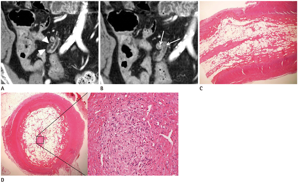
Fig. 1 Computed tomography (CT) findings and pathological images of fibrous obliteration in the appendix of a 63-year-old man. A. Coronal CT scan shows borderline dilatation of the appendix filled with a fatty component (-33 Hounsfield unit). A focal wall defect in the distal portion of the appendix and an adjacent mild fatty infiltration (arrowheads) is visible. B. Coronal CT scan shows the linear-enhancing core of the appendix (arrows). C, D. Photomicrographs of a histopathological specimen in longitudinal (C) and cross-sectional (D) view of the distal appendix [hematoxylin and eosin stain, × 1.25 (C, D-left), × 200 (D-right)] show adipocyte and fibroblast proliferation filling the appendiceal lumen and focal destruction of the mucosa (or ragged mucosa). Several arterioles are visible in the center. The suspicious focal wall defect on the CT scan was not visible in the histopathological specimen.
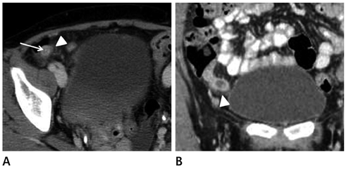
Fig. 2 Computed tomography (CT) findings of fibrous obliteration in the appendix of a 43-year-old woman. A. Axial CT scan shows periappendiceal infiltration (arrowhead) and a linear-enhancing core structure (arrow). B. Coronal CT scan shows periappendiceal infiltration (arrowhead). Note the thickened appendiceal wall and dilated lumen.
Reference
-
1. Misdraji J, Graeme-Cook FM. Miscellaneous conditions of the appendix. Semin Diagn Pathol. 2004; 21:151–163.2. Michalany J, Galindo W. Classification of neuromas of the appendix. Beitr Pathol. 1973; 150:213–228.3. Noffsinger A, Fenoglio-Preiser CM, Maru D, Gilinsky N. Gastrointestinal diseases: atlas of nontumor pathology. Washington DC: American Registry of Pathology;2007. p. 633–634.4. Stanley MW, Cherwitz D, Hagen K, Snover DC. Neuromas of the appendix. A light-microscopic, immunohistochemical and electron-microscopic study of 20 cases. Am J Surg Pathol. 1986; 10:801–815.5. Olsen BS, Holck S. Neurogenous hyperplasia leading to appendiceal obliteration: an immunohistochemical study of 237 cases. Histopathology. 1987; 11:843–849.6. Jones B, Fishman EK, Hamilton SR, Rubesin SE, Bayless TM, Cameron JC, et al. Submucosal accumulation of fat in inflammatory bowel disease: CT/pathologic correlation. J Comput Assist Tomogr. 1986; 10:759–763.7. Muldowney SM, Balfe DM, Hammerman A, Wick MR. "Acute" fat deposition in bowel wall submucosa: CT appearance. J Comput Assist Tomogr. 1995; 19:390–393.8. Raman SS, Lu DS, Kadell BM, Vodopich DJ, Sayre J, Cryer H. Accuracy of nonfocused helical CT for the diagnosis of acute appendicitis: a 5-year review. AJR Am J Roentgenol. 2002; 178:1319–1325.9. Rhea JT, Halpern EF, Ptak T, Lawrason JN, Sacknoff R, Novelline RA. The status of appendiceal CT in an urban medical center 5 years after its introduction: experience with 753 patients. AJR Am J Roentgenol. 2005; 184:1802–1808.10. Checkoff JL, Wechsler RJ, Nazarian LN. Chronic inflammatory appendiceal conditions that mimic acute appendicitis on helical CT. AJR Am J Roentgenol. 2002; 179:731–734.11. Crabbe MM, Norwood SH, Robertson HD, Silva JS. Recurrent and chronic appendicitis. Surg Gynecol Obstet. 1986; 163:11–13.
- Full Text Links
-
- Actions
-
Cited
- CITED
-
- Close
- Share
-
- Similar articles
-
- Pathologic Analysis of 2159 Cases of Appendix
- Endometriosis of the Appendix
- Tuberculosis of the Appendix Associated with as Acute Suppurative Inflammatory Reaction: A case report
- A Case of Incidental Chronic Appendicitis: Appendiceal Wall Thickening in an Asymptomatic Patient
- Preoperative Diagnosis of Diverticulitis of the Vermiform Appendix by Ultrasonography
