J Korean Soc Radiol.
2015 Apr;72(4):295-299. 10.3348/jksr.2015.72.4.295.
Bilateral Presentation of Pleural Desmoplastic Small Round Cell Tumors: A Case Report
- Affiliations
-
- 1Department of Radiology, Soonchunhyang University College of Medicine, Bucheon Hospital, Bucheon, Korea. jspark@schmc.ac.kr
- 2Department of Pathology, Soonchunhyang University College of Medicine, Bucheon Hospital, Bucheon, Korea.
- KMID: 1941762
- DOI: http://doi.org/10.3348/jksr.2015.72.4.295
Abstract
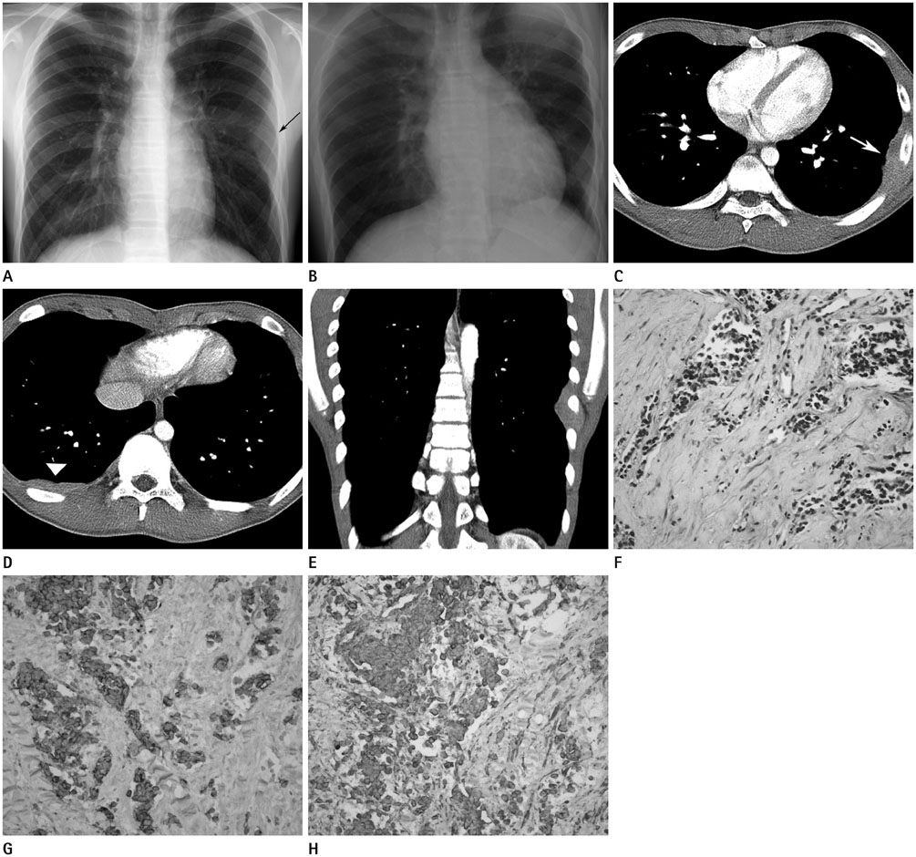
- Desmoplastic small round cell tumor (DSRCT) is a highly aggressive malignant small cell neoplasm occurring mainly in the abdominal cavity, but it is extremely rare in the pleura. In this case, a 15-year-old male presented with a 1-month history of left chest pain. Chest radiographs revealed pleural thickening in the left hemithorax and chest computed tomography showed multifocal pleural thickening with enhancement in both hemithoraces. A needle biopsy of the left pleural lesion was performed and the final diagnosis was DSRCT of the pleura. We report this unusual case arising from the pleura bilaterally. The pleural involvement of this tumor supports the hypothesis that it typically occurs in mesothelial-lined surfaces.
MeSH Terms
Figure
Reference
-
1. Gerald WL, Rosai J. Case 2. Desmoplastic small cell tumor with divergent differentiation. Pediatr Pathol. 1989; 9:177–118.2. Biswas G, Laskar S, Banavali SD, Gujral S, Kurkure PA, Muckaden M, et al. Desmoplastic small round cell tumor: extra abdominal and abdominal presentations and the results of treatment. Indian J Cancer. 2005; 42:78–84.3. Parkash V, Gerald WL, Parma A, Miettinen M, Rosai J. Desmoplastic small round cell tumor of the pleura. Am J Surg Pathol. 1995; 19:659–665.4. Ostoros G, Orosz Z, Kovács G, Soltész I. Desmoplastic small round cell tumour of the pleura: a case report with unusual follow-up. Lung Cancer. 2002; 36:333–336.5. Lal DR, Su WT, Wolden SL, Loh KC, Modak S, La Quaglia MP. Results of multimodal treatment for desmoplastic small round cell tumors. J Pediatr Surg. 2005; 40:251–255.6. Kis B, O'Regan KN, Agoston A, Javery O, Jagannathan J, Ramaiya NH. Imaging of desmoplastic small round cell tumour in adults. Br J Radiol. 2012; 85:187–192.7. Xu Q, Xu K, Yang C, Zhang X, Meng Y, Quan Q. Askin tumor: four case reports and a review of the literature. Cancer Imaging. 2011; 11:184–188.8. Dynes MC, White EM, Fry WA, Ghahremani GG. Imaging manifestations of pleural tumors. Radiographics. 1992; 12:1191–1201.9. Li H, Smolen GA, Beers LF, Xia L, Gerald W, Wang J, et al. Adenosine transporter ENT4 is a direct target of EWS/WT1 translocation product and is highly expressed in desmoplastic small round cell tumor. PLoS One. 2008; 3:e2353.10. Modak S, Gerald W, Cheung NK. Disialoganglioside GD2 and a novel tumor antigen: potential targets for immunotherapy of desmoplastic small round cell tumor. Med Pediatr Oncol. 2002; 39:547–551.
- Full Text Links
- Actions
-
Cited
- CITED
-
- Close
- Share
- Similar articles
-
- A case of pelvic desmoplastic small round cell tumor in a old aged woman
- A case of peritoneal desmoplastic small round cell tumor
- A Case of Peritoneal Desmoplastic Small Round Cell Tumor which involved both ovaries
- Desmoplastic Small Round Cell Tumor: A Case Report
- Intra-abdominal Desmoplastic Small Round Cell Tumor Diagnosed by Lymph Node Biopsy: A case report


