Korean J Phys Anthropol.
2014 Jun;27(2):65-70. 10.11637/kjpa.2014.27.2.65.
Morphometric Study of the Retromolar Triangle and Foramen in Korean Mandibles
- Affiliations
-
- 1Department of Oral Anatomy, School of Dentistry, Pusan National University, Korea. kwakhh@pusan.ac.kr
- 2Department of Physiology, College of Medicine, Seonam University, Korea.
- KMID: 1917125
- DOI: http://doi.org/10.11637/kjpa.2014.27.2.65
Abstract
- It has been known that the retromolar foramen is a rare anatomic variation observed in the retromolar triangle, a small triangular shaped region posterior to the mandibular third molar. Due to the neurovascular bundle passing through the retromolar foramen, this anatomical structure must be kept in mind during surgical approaches regarding the retromolar area and mandible. Therefore, the authors investigated the morphology of retromolar triangle and the existence and location of retromolar foramen in Korean. And these results were compared with that of other races. We used 308 sides of 154 Korean dry mandibles, unknown gender and age. The retromolar triangle presented predominantly a triangular shape (84.1%), and the maximum height and width were 13.7 mm and 7.1 mm, respectively. In 144 of the 308 sides, the retromolar foramen was observed (46.8%). The existence of the retromolar foramen was seen the same frequency in both sides, and based on a midsagittal line of the retromolar triangle, the retromolar foramen located in more buccal side (75%) than lingual side. The mean distance between the retromolar foramen and the distal edge of the last tooth were found to be 10.3 mm and 6.9 mm, respectively for the second and third molars. According to the present study, the northeast Asians including Korean population show the highest rate of the incidence of the retromolar foramen than other races. The findings suggest that practitioners should take the retromolar foramen into account in surgical procedures involving the retromolar area to protect the patient from the complications such as bleeding or nerve damage.
Keyword
MeSH Terms
Figure
-
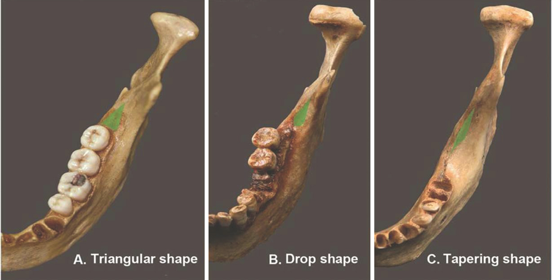
Fig. 1. Classifications of the RMT according to the shape. (A) Triangular shape (84.1%), (B) Drop shape(9.3%), and (C) Tapering shape (6.6%).
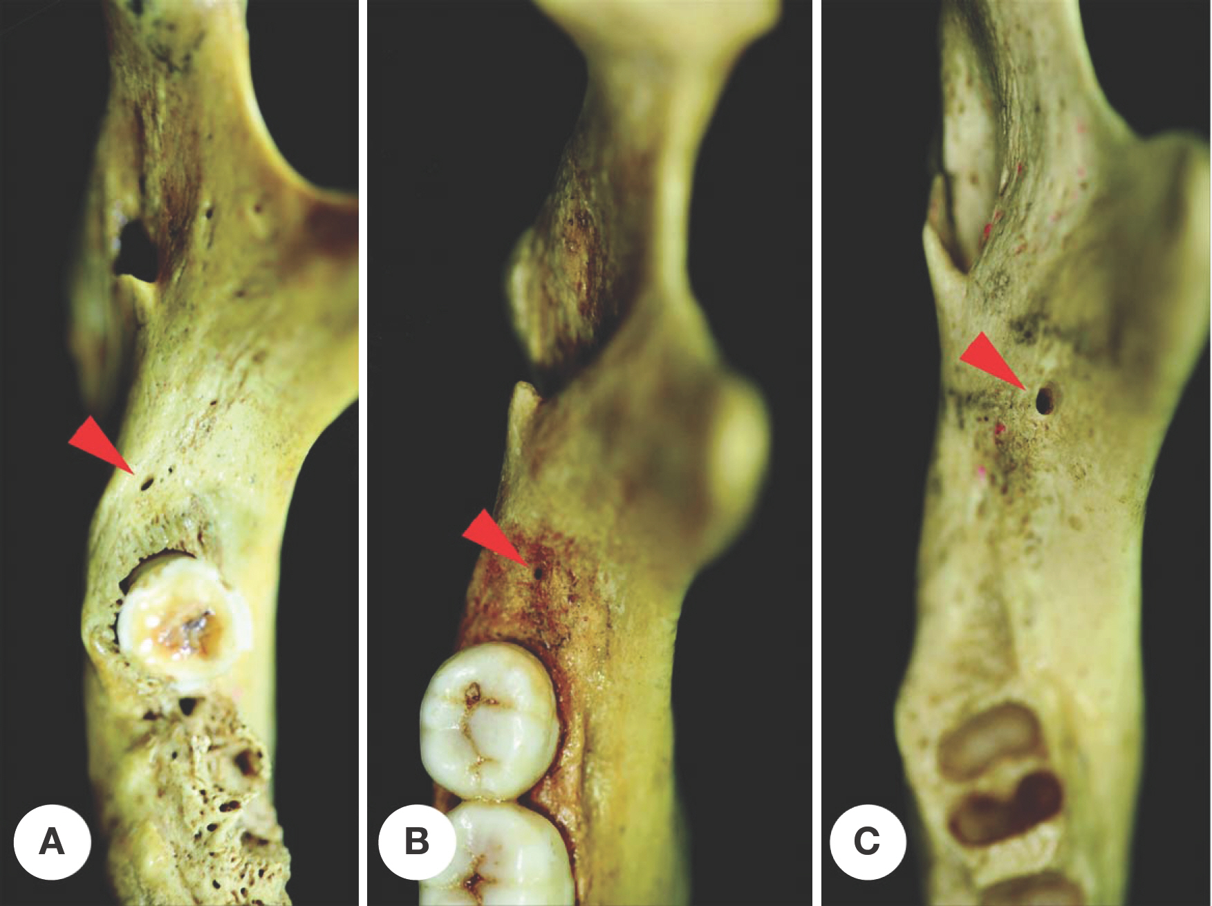
Fig. 2. Photographs showing the RMF(arrowheads). (A) RMF located in the RMT and on the lingual side of the midsagittal line of RMT,(B) RMF located in the RMT and on the midsagittal line of RMT, and (C) RMF located out of the RMT and on the buccal side of the midsagittal line of RMT.
Reference
-
References
1. Bilecenoglu B, Tuncer N. Clinical and anatomical study of retromolar foramen and canal. J Oral Maxillofac Surg. 2006; 64:1493–7.
Article2. Schejtman R, Devoto FCH, Arias NH. The origin and distribution of the elements of the human mandibular retromolar canal. Arch Oral Biol. 1967; 12:1261–7.
Article3. von Arx T, Hänni A, Sendi P, Buser D, Bornstein MM. Radiographic study of the mandibular retromolar canal: an anatomic structure with clinical importance. J Endod. 2011; 37:1630–5.
Article4. Kawai T, Asaumi R, Sato I, Kumazawa Y, Yosue T. Observation of the retromolar foramen and canal of the mandible: a CBCT and macroscopic study. Oral Radiol. 2012; 28:10–4.
Article5. Ossenberg NS. Temporal crest canal: case report and statistics on a rare mandibular variant. Oral Surg Oral Med Oral Pathol. 1986; 62:10–2.
Article6. Hu KS, Koh KS, Park KK, Kang MK, Chung IH, Kim HJ. Nonmetric traits of Korean mandibles. Korean J Phys An-throp. 2000; 13:161–72. .(article in Korean).
Article7. Naitoh M, Nakahara K, Suenaga Y, Gotoh K, Kondo S, Ariji E. Variations of the bony canal in the mandibular ramus using cone-beam computed tomography. Oral Radiol. 2010; 26:36–40.
Article8. Suazo GI, Cantin LM, Lopez FB, Valenzuela UV, Valenzuela RR. Morphometric study of the retromolar triangle. Int J Odontostomat. 2007; 1:129–32.9. Kodera H, Hashimoto I. A case of mandibular retromolar canal: elements of nerves and arteries in this canal. Kaibo-gaku Zasshi. 1995; 70:23–30. .(article in Japanese).10. Szycik V, Dijakiewicz M, Zienkiewicz J, Pawlowska A. Bio-Oss: Its use as an extension of indications for implant den-tistry. Ann Acad Med Ged. 2002; 32:151–9.11. Suazo-Galdames IC, Cantín-López MG, Zavando-Matamala DA. Inferior alveolar nerve block anesthesia via the retromolar triangle, an alternative for patients with blood dyscrasias. Med Oral Patol Oral Cir Bucal. 2008; 13(1):E43–7.12. Sawyer DR, Kiely ML. Retromolar foramen: a mandibular variant important to dentistry. Ann Dent. 1991; 50:16–8.13. Muller H. Caracteres non-metriques du squelette de la tete chez les populations medievales de Thoiry(Ain, France) et de Bavois (Vaud, Suisse). Arch Suisses Anthropol Gen. 1977; 41:123–64.14. Ossenberg NS. Retromolar foramen of the human mandible. Am J Phys Anthropol. 1987; 73:119–28.
Article15. Pyle MA, Jasinevicius TR, Lalumandier JA, Kohrs KJ, Sawyer DR. Prevalence and implications of accessory retromolar foramina in clinical dentistry. General Dent. 1999; 47:500–3.16. Löfgren AB. Foramina retromolaria mandibulae. A study on human skulls of nutrient foramina situated in the mandibular retromolar fossa. Odont Tidskr. 1957; 65:552–70.17. Ahmed WN, Bhat JG, Narayana K, Nayak UA, Devaiah BA. The retromolar foramen and canal in south Indian dry man-dibles. European J Anat. 2002; 6:141–6.
- Full Text Links
-
- Actions
-
Cited
- CITED
-
- Close
- Share
-
- Similar articles
-
- Morphometric study on mandibular foramen and incidence of accessory mandibular foramen in mandibles of south Indian population and its clinical implications in inferior alveolar nerve block
- Morphological study of the mental spine, lingual foramen and nutrient foramen and innominate foramen in Korean mandibles
- Imaging of Jaw with Dental CT Software Program Normal Anatomy
- Anatomical Study on the Location of the Mental Foramen in Adult Korean Mandibles
- A Morphometric Study on the Mandibular Foramen and the Lingula in Korean
