Korean J Urol.
2007 Jun;48(6):663-666. 10.4111/kju.2007.48.6.663.
Pelvic Schwannoma Causing Recurrent Acute Urinary Retention
- Affiliations
-
- 1Department of Urology, College of Medicine, Hanyang University, Seoul, Korea. moonuro@hanyang.ac.kr
- 2Department of Pathology, College of Medicine, Hanyang University, Seoul, Korea.
- KMID: 1885552
- DOI: http://doi.org/10.4111/kju.2007.48.6.663
Abstract
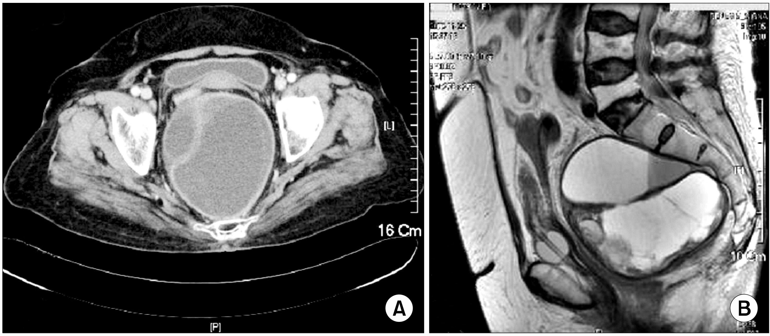
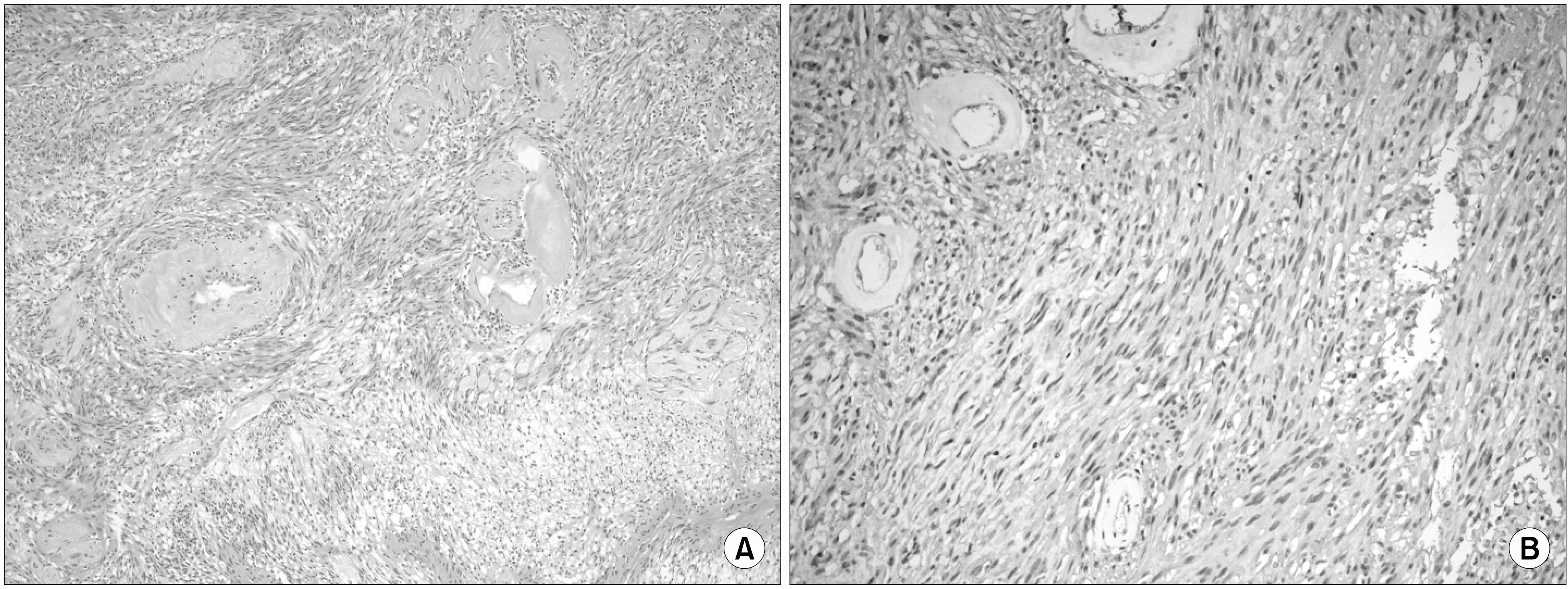
- Schwannoma is a tumor that arises from neural sheath Schwann cells of peripheral nerves. Schwannoma is mostly solitary, except when it occurs in association with Von Recklinghausen's disease. Solitary schwannoma can occur in association with a nerve anywhere within the body, but rarely occurs in the pelvis. Microscopically, the tumors can be divided into hypercellular bundles of spindle-shaped cells (Antoni A area) and areas of lower cellularity, with loose myxomatous arrangement of cells and fibers (Antoni B area). Complete resection of pelvic schwannoma is a curative treatment. We report a case of benign presacral cystic schwannoma that caused recurrent acute urinary retention in a 79-year-old woman, along with a review of the literature.
Keyword
MeSH Terms
Figure
Reference
-
References
1. Patocskai EJ, Tabatabaian M, Thomas MJ. Cellular schwannoma: a rare presacral tumour. Can J Surg. 2002; 45:141–4.2. Goh BK, Tan YM, Chung YF, Chow PK, Ooi LL, Wong WK. Retroperitoneal schwannoma. Am J Surg. 2006; 192:14–8.
Article3. Jung JH, Jung HK, Kam SC, Choi SM, Hyun JS, Jung KH, et al. Schwannoma originated from obturator nerve of pelvic cavity in patient with urinary frequency. Korean J Urol. 2005; 46:992–4.4. Yoon HC, Chung CY, Chun SJ, Park KM, Park HS, Rho J, et al. A case of pelvic neurilemoma. Korean J Urol. 2000; 41:907–9.5. Oh KS, Ha SI, Lee HS, Lee JS, Kwak SS, Yun SH. Giant benign schwannoma involving sacral bone. J Korean Neurosurg Soc. 2001; 30:509–13.6. Hughes MJ, Thomas JM, Fisher C, Moskovic EC. Imaging features of retroperitoneal and pelvic schwannomas. Clin Radiol. 2005; 60:886–93.
Article7. Dede M, Yagci G, Yenen MC, Gorgulu S, Deveci MS, Cetiner S, et al. Retroperitoneal benign schwannoma: report of three cases and analysis of clinicoradiologic findings. Tohoku J Exp Med. 2003; 200:93–7.
Article8. Daneshmand S, Youssefzadeh D, Chamie K, Boswell W, Wu N, Stein JP, et al. Benign retroperitoneal schwannoma: a case series and review of the literature. Urology. 2003; 62:993–7.
Article9. Rosai J. Soft tissues. Rosai J, editor. Ackerman's surgical pathology. 8th ed.St. Louis: Mosby;1996. p. 2041–53.10. Hwang TS, Park SH, Ham EK. Histopathologic and immunohistochemical observation on malignant schwannoma. Korean J Pathol. 1990; 24:446–55.


