Multiple absences of the branches of abdominal aorta with congenital absence of the portal vein, unilateral adrenal agenesis and persistent ductus arteriosus in a female cadaver
- Affiliations
-
- 1Department of Anatomy and Pathobiology, School of Medicine, North Khorasan University of Medical Sciences, Bojnurd, Iran. Shahahmadpour@gmail.com
- KMID: 1882608
- DOI: http://doi.org/10.5115/acb.2014.47.4.274
Abstract
- We report on an extremely rare case of multiple absences of the branches of abdominal aorta with congenital absence of the portal vein, unilateral adrenal agenesis and persistent ductus arteriosus in an adult female cadaver. Specifically, instead of celiac trunk, superior and inferior mesenteric arteries, solely a single arterial trunk aroused from the anterior aspect of abdominal aorta, inferior phrenic and ovarian arteries were absent in both sides. Left kidneys drained by two veins. There were not superior, splenic and mesenteric veins, while left renal vein received an additional vein, which run downward and drained primarily all parts of digestive tract and its associated glands (portal vein did not exist). Right adrenal gland was absent. To the best of our knowledge, it is the only reported case with such widespread anomalies. We think the importance of this case is beyond the surgical consideration and needs more profound developmental studies.
MeSH Terms
Figure
-
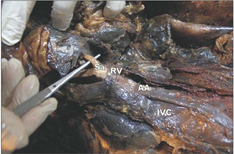
Fig. 1 Abdominal cavity right side view, small and large intestine have been removed upward and abdominal aorta (AA) and its single arterial branch (Sa) exposed. The Sa enters the mesentery. Left renal vein (RV) runs horizontally to right and drain to inferior vena cava (IVC).

Fig. 2 Aorta has been dissected and removed with intestines and kidneys. Thorasic aorta (ThA), abdominal aorta (AA), right and left kidneys (LK and RK). AA and its branches (two renal arteries and single branch to digestive tract). Celiac trunk, phrenic, superior and inferior mesenteric and ovarian arteries were absent.

Fig. 3 Left kidney drains by two veins, anterior left reneal vein (LRVa) passes anterior to abdominal aorta (AA) and another vein, posterior left renal vein (LRVp) run posterior to AA and collectively drain to inferior vena cava.
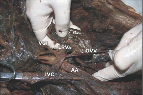
Fig. 4 Anterior left renal vein (LRVa) receives ovarian vein (OVV) and a single vein form gastrointestinal tract (VGI). AA, abdominal aorta; IVC, inferior vena cava.
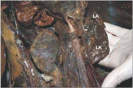
Fig. 5 Left adrenal (LA) gland and left kidney (LK). Adrenal gland was absent in right side.
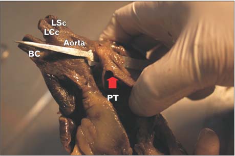
Fig. 6 Persistent ductus arteriosus. Pulmonary trunk (PT) connects to aorta by persistent ductus arteriosus (arrow). BC, brachiocephalic trunk; LCc, left common carotid; LSc, left subsabclavian.
Cited by 1 articles
-
Prevalence and clinical relevance of the anatomical variations of suprarenal arteries: a review
Ananya Priya, Ravi Kant Narayan, Sanjib Kumar Ghosh
Anat Cell Biol. 2022;55(1):28-39. doi: 10.5115/acb.21.211.
Reference
-
1. Dogan NU, Fazliogullari Z, Yilmaz MT, Uysal II, Cicekcibasi AE, Ulusoy M, Gunaslan P. A complex variation of the parietal and visceral branches of the abdominal aorta. Int J Morphol. 2011; 29:90–93.2. Songür A, Toktaş M, Alkoç O, Acar T, Uzun I, Baş O, Özen OA. Abdominal aorta and its branches: morphometry. Variations in autopsy cases. Eur J Gen Med. 2010; 7:321–325.3. Shivarama CH, Bhat S, Shetty RK, Avadhani R. Multiple variations of branches of abdominal aorta: a case study. Nitte Univ J Health Sci. 2012; 2:48–50.4. Kalthur SG, Sarda R, Bankar M. Multiple vascular variations of abdominal vessels in a male cadaver: embryological perspective and clinical importance. J Morphol Sci. 2011; 28:152–156.5. Morse SS, Taylor KJ, Strauss EB, Ramirez E, Seashore JH. Congenital absence of the portal vein in oculoauriculovertebral dysplasia (Goldenhar syndrome). Pediatr Radiol. 1986; 16:437–439.6. Venkat-Raman N, Murphy KW, Ghaus K, Teoh TG, Higham JM, Carvalho JS. Congenital absence of portal vein in the fetus: a case report. Ultrasound Obstet Gynecol. 2001; 17:71–75.7. Northrup M, Mendez-Castillo A, Sethi Y, Churchill R. Congenital absence of the portal vein with an intrahepatic inferior vena cava branch showing hepatopetal flow. J Ultrasound Med. 2002; 21:569–572.8. Gupta A, Singal R, Singh D. Variations of gonadal artery: embryological basis and clinical significance. Int J Biol Med Res. 2011; 2:1006–1010.9. Kasindye GU, Mwasunga AS, Fabian FM. Bilateral absence of ovarian artery in a Tanzanian female cadaver: a rare variation. Int J Anat Var. 2012; 5:73–75.10. Lipshutz B. A composite study of the coeliac axis artery. Ann Surg. 1917; 65:159–169.11. Chen H, Yano R, Emura S, Shoumura S. Anatomic variation of the celiac trunk with special reference to hepatic artery patterns. Ann Anat. 2009; 191:399–407.12. Morita M. Reports and conception of three anomalous cases on the area of the celiac and the superior mesenteric arteries. Igaku Kenkyu. 1935; 9:1993–2006.13. Yi SQ, Terayama H, Naito M, Hirai S, Alimujang S, Yi N, Tanaka S, Itoh M. Absence of the celiac trunk: case report and review of the literature. Clin Anat. 2008; 21:283–286.14. Matusz P, Miclaus GD, Ples H, Tubbs RS, Loukas M. Absence of the celiac trunk: case report using MDCT angiography. Surg Radiol Anat. 2012; 34:959–963.15. Wu Y, Peng W, Wu H, Chen G, Zhu J, Xing C. Absence of the superior mesenteric artery in an adult and a new classification method for superior-inferior mesenteric arterial variations. Surg Radiol Anat. 2014; 36:511–515.
- Full Text Links
-
- Actions
-
Cited
- CITED
-
- Close
- Share
-
- Similar articles
-
- Congenital absence of ductus arteriosus: an autopsy case
- A case of coaretation of the aorta associated with the patent ductus arteriosus and the persistent leftsuperior vena cava
- Lobar Agenesis of the Liver'Imaging Findings
- Unilateral Renal Agenesis With Spontaneous Adrenal Hemorrhage: One Case
- A Case of Interruption of Aorta with Patent Ductus Arteriosus
