Anat Cell Biol.
2014 Dec;47(4):267-270. 10.5115/acb.2014.47.4.267.
Unilateral ureteric stone associated with gross hydronephrosis and kidney shrinkage: a cadaveric report
- Affiliations
-
- 1Department of Anatomy, Yong Loo Lin School of Medicine, National University of Singapore, Singapore, Singapore. antoi@nus.edu.sg antngyk@nus.edu.sg
- KMID: 1882606
- DOI: http://doi.org/10.5115/acb.2014.47.4.267
Abstract
- Ureteric stones are a common cause of obstruction of the urinary tract, usually presenting with characteristic signs and symptoms, such as acute ureteric colic and hematuria. Occasionally, stones may present with non-specific symptoms such as low back pain and remain unidentified, leading to stone growth, chronic ureteric obstruction and complications such as hydronephrosis and renal damage. Here, we report a large ureteric stone in a cadaver with complete obstruction at the left ureterovesical junction, resulting in severe dilatation of the left ureter and renal pelvis.
Keyword
MeSH Terms
Figure
-
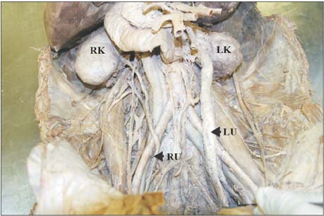
Fig. 1 Photograph showing the severely dilated left ureter as well as shrunken nodular appearance of the left kidney. Although the left ureter was completely obstructed, the right kidney does not seem to have any compensatory enlargement. LK, left kidney; LU, left ureter; RK, right kidney; RU, right ureter.

Fig. 2 Macroscopic view of the huge, dark-coloured ureteric stone (25mm×11 mm) lodged at the left ureterovesical junction.

Fig. 3 Sagittal section view of the left kidney demonstrating a dilated renal pelvis due to prolonged obstruction. DRP, dilated renal pelvis; HU, hydronephrotic ureter.
Reference
-
1. Teichman JM. Clinical practice. Acute renal colic from ureteral calculus. N Engl J Med. 2004; 350:684–693.2. Hall PM. Nephrolithiasis: treatment, causes, and prevention. Cleve Clin J Med. 2009; 76:583–591.3. Chung C, Stern PJ, Dufton J. Urolithiasis presenting as right flank pain: a case report. J Can Chiropr Assoc. 2013; 57:69–75.4. Wells KA. Nephrolithiasis with unusual initial symptoms. J Manipulative Physiol Ther. 2000; 23:196–201.5. Wimpissinger F, Springer C, Kurtaran A, Stackl W, Turk C. Functional aspects of silent ureteral stones investigated with MAG-3 renal scintigraphy. BMC Urol. 2014; 14:3.6. Colella J, Kochis E, Galli B, Munver R. Urolithiasis/nephrolithiasis: whats it all about? Urol Nurs. 2005; 25:427–448. 4754497. Wimpissinger F, Türk C, Kheyfets O, Stackl W. The silence of the stones: asymptomatic ureteral calculi. J Urol. 2007; 178(4 Pt 1):1341–1344.8. Shimada K, Matsumoto F, Tohda A, Ueda M. Histological study of fetal kidney with urethral obstruction and vesicoureteral reflux: a consideration on the etiology of congenital reflux nephropathy. Int J Urol. 2003; 10:518–524.9. Huland H, Gonnermann D. Pathophysiology of hydronephrotic atrophy: the cause and role of active preglomerular vasoconstriction. Urol Int. 1983; 38:193–198.10. Marchini GS, Vicentini FC, Mazzucchi E, Brito A, Ebaid G, Srougi M. Silent ureteral stones: impact on kidney function: can treatment of silent ureteral stones preserve kidney function? Urology. 2012; 79:304–308.11. Zulkifli MZ, Ho CC, Goh EH, Praveen S, Das S. Ureteric stone in the presence of existing backache: lessons to learn. Clin Ter. 2012; 163:23–25.12. Vaidyanathan S, Singh G, Soni BM, Hughes P, Watt JW, Dundas S, Sett P, Parsons KF. Silent hydronephrosis/pyonephrosis due to upper urinary tract calculi in spinal cord injury patients. Spinal Cord. 2000; 38:661–668.13. Wolcott CC. An atypical case of nephrolithiasis with transient remission of symptoms following spinal manipulation. J Chiropr Med. 2010; 9:69–72.
- Full Text Links
-
- Actions
-
Cited
- CITED
-
- Close
- Share
-
- Similar articles
-
- Three Cases of Horseshoe Kidneys with Complications.
- A Case of Horseshoe Kidney with Single Renal Stone and Hydronephrosis
- Giant Hydronephrosis Combined with Ureteral Stone
- Two Cases of Unilateral Renal Agenesis
- Unilateral Ureteric Entrapment within the Sacroiliac Joint Causing Unilateral Hydroureteronephrosis
