J Periodontal Implant Sci.
2014 Feb;44(1):8-12. 10.5051/jpis.2014.44.1.8.
Survival of 352 titanium implants placed in 181 patients: a 4-year multicenter field study
- Affiliations
-
- 1Department of Periodontology, Institute of Oral Biology, Kyung Hee University School of Dentistry, Seoul, Korea. chungjh@khu.ac.kr
- 2Division of Periodontology, Department of Dentistry, Inha University School of Medicine, Incheon, Korea.
- 3Department of Periodontology, Kyung Hee University School of Dentistry, Seoul, Korea.
- KMID: 1845872
- DOI: http://doi.org/10.5051/jpis.2014.44.1.8
Abstract
- PURPOSE
The aim of this retrospective chart review was to evaluate the four-year survival rate of a titanium implant system.
METHODS
A total of 352 sand-blasted, thermally acid-etched titanium implants were inserted into 181 partially or completely edentulous patients. Their cumulative survival rate was evaluated retrospectively. Associated factors, such as the implant distribution and treatment type were included in the evaluation.
RESULTS
The implants were equally distributed between the maxilla (52.3%) and the mandible (47.7%). 48 implants (13.6%) were placed in the anterior region and 304 implants (86.4%) in the posterior region. The majority of the implants were inserted into bone of type II and III quality (89.8%) and volume (quantity B and C, 87.2%). Most of the implants (70.7%) were restored as single crowns; 28.7% supported a bridge construction and 0.6% a full denture. Only one implant failed, resulting in a four-year cumulative survival rate of 99.7%.
CONCLUSIONS
The implant system showed an excellent four-year survival rate. It proved to be a safe and predictable means for restoration of the dentition in partially or completely edentulous patients.
Keyword
MeSH Terms
Figure
-

Figure 1 Number of implants inserted by the 1 (nonsubmerged) and 2 stage (submerged) surgical protocols. Mx. Ant.: anterior maxilla, Mx. Post.: posterior maxilla, Mn. Ant.: anterior mandible, Mn. Post.: posterior mandible.
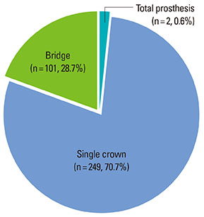
Figure 2 Implant restoration types.
Reference
-
1. Adell R, Lekholm U, Rockler B, Branemark PI. A 15-year study of osseointegrated implants in the treatment of the edentulous jaw. Int J Oral Surg. 1981; 10:387–416.
Article2. Henry PJ, Laney WR, Jemt T, Harris D, Krogh PH, Polizzi G, et al. Osseointegrated implants for single-tooth replacement: a prospective 5-year multicenter study. Int J Oral Maxillofac Implants. 1996; 11:450–455.3. Lekholm U, Gunne J, Henry P, Higuchi K, Linden U, Bergstrom C, et al. Survival of the Brånemark implant in partially edentulous jaws: a 10-year prospective multicenter study. Int J Oral Maxillofac Implants. 1999; 14:639–645.4. Cochran D. Implant therapy I. Ann Periodontol. 1996; 1:707–791.
Article5. Martinez H, Davarpanah M, Missika P, Celletti R, Lazzara R. Optimal implant stabilization in low density bone. Clin Oral Implants Res. 2001; 12:423–432.
Article6. Meredith N. Assessment of implant stability as a prognostic determinant. Int J Prosthodont. 1998; 11:491–501.7. Barewal RM, Oates TW, Meredith N, Cochran DL. Resonance frequency measurement of implant stability in vivo on implants with a sandblasted and acid-etched surface. Int J Oral Maxillofac Implants. 2003; 18:641–651.8. Buser D, Nydegger T, Oxland T, Cochran DL, Schenk RK, Hirt HP, et al. Interface shear strength of titanium implants with a sandblasted and acid-etched surface: a biomechanical study in the maxilla of miniature pigs. J Biomed Mater Res. 1999; 45:75–83.
Article9. Cochran DL. A comparison of endosseous dental implant surfaces. J Periodontol. 1999; 70:1523–1539.
Article10. Cutler SJ, Ederer F. Maximum utilization of the life table method in analyzing survival. J Chronic Dis. 1958; 8:699–712.
Article11. Lekholm U, Zarb GA. Patient selection and preparation. In : Branemark PI, Zarb GA, Albrektsson T, editors. Tissue-integrated prostheses: osseointegration in clinical dentistry. . Chicago: Quintessence;1985. p. 199–209.12. Albrektsson T, Zarb GA. Current interpretations of the osseointegrated response: clinical significance. Int J Prosthodont. 1993; 6:95–105.13. Buser D, Mericske-Stern R, Bernard JP, Behneke A, Behneke N, Hirt HP, et al. Long-term evaluation of non-submerged ITI implants. Part 1: 8-year life table analysis of a prospective multi-center study with 2359 implants. Clin Oral Implants Res. 1997; 8:161–172.
Article14. Krennmair G, Seemann R, Schmidinger S, Ewers R, Piehslinger E. Clinical outcome of root-shaped dental implants of various diameters: 5-year results. Int J Oral Maxillofac Implants. 2010; 25:357–366.15. Cochran D, Oates T, Morton D, Jones A, Buser D, Peters F. Clinical field trial examining an implant with a sand-blasted, acid-etched surface. J Periodontol. 2007; 78:974–982.
Article16. Albrektsson T, Wennerberg A. Oral implant surfaces: Part 1: review focusing on topographic and chemical properties of different surfaces and in vivo responses to them. Int J Prosthodont. 2004; 17:536–543.17. Lossdörfer S, Schwartz Z, Wang L, Lohmann CH, Turner JD, Wieland M, et al. Microrough implant surface topographies increase osteogenesis by reducing osteoclast formation and activity. J Biomed Mater Res A. 2004; 70:361–369.
Article18. Buser D, Schenk RK, Steinemann S, Fiorellini JP, Fox CH, Stich H. Influence of surface characteristics on bone integration of titanium implants: a histomorphometric study in miniature pigs. J Biomed Mater Res. 1991; 25:889–902.
Article19. Wennerberg A, Ektessabi A, Albrektsson T, Johansson C, Andersson B. A 1-year follow-up of implants of differing surface roughness placed in rabbit bone. Int J Oral Maxillofac Implants. 1997; 12:486–494.20. Lazzara RJ, Testori T, Trisi P, Porter SS, Weinstein RL. A human histologic analysis of osseotite and machined surfaces using implants with 2 opposing surfaces. Int J Periodontics Restorative Dent. 1999; 19:117–129.
- Full Text Links
-
- Actions
-
Cited
- CITED
-
- Close
- Share
-
- Similar articles
-
- Retrospective multicenter study of CSM endosseous dental implant
- A comparative clinical study on oxidized titanium implants and sandblasted large-grit acid etched implants in soft bone
- The skeletal cortical anchorage using titanium microscrew implants
- ULTRASTRUCTURAL INVESTIGATIONS OF THE INTERFACE BETWEEN CULTURED PERIODONTAL LIGAMENT CELLS AND TITANIUM
- A histologic comparative study of loaded and unloaded titanium implants
