Tracheobronchial Polyps Following Thermal Inhalation Injury
- Affiliations
-
- 1Division of Pulmonary and Critical Care Medicine, Department of Medicine, Samsung Medical Center, Sungkyunkwan University School of Medicine, Seoul, Korea. kjeon@skku.edu
- 2Division of Pulmonary and Critical Care Medicine, Department of Internal Medicine, The Armed Forces Capital Hospital, Seongnam, Korea.
- KMID: 1842970
- DOI: http://doi.org/10.4046/trd.2014.76.5.237
Abstract
- The early pulmonary consequences of inhalation injury are well documented; however, little is known about delayed pulmonary complications following thermal inhalation injury. Although thermal injury below the vocal cords is rare because of effective heat dissipation in the upper airway, inflammatory endobronchial polyps have previously been reported as a delayed complication associated with inhalation injury. We report an extraordinary case of tracheobronchial polyps in patients with smoke inhalation injury. This report shows the delayed development and natural course of tracheobronchial polyps following thermal injury.
Keyword
MeSH Terms
Figure
-
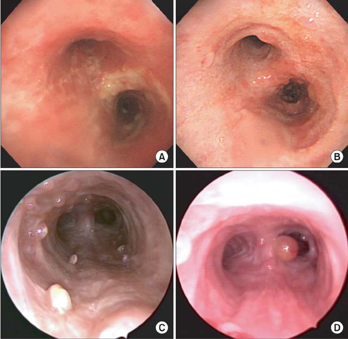
Figure 1 A 21-year-old man with inhalation burn in the tracheobronchial wall. (A) Bronchoscopy showed edematous and hyperemic mucosal changes in the tracheobronchial wall at 2 weeks after the inhalation injury. (B) One month after the inhalation injury, numerous pale polypoid lesions were found in the tracheobronchial tree. (C) Three months after the initial injury, endobronchial polyposis had not changed, and background mucosa in the trachea appeared to be fibrotic in nature. (D) Six months after the initial injury, the lesions were slightly reduced with fibrotic change at the trachea, but an additional exophytic polyp was seen at the right side of the carina.

Figure 2 A 21-year-old man with inhalation burn in the tracheobronchial tree. Microscopic findings of the tracheobronchial polyps revealed granulomatous inflammation (A, H&E stain, ×100; B, H&E stain, ×200).
Reference
-
1. Heimbach DM, Waeckerle JF. Inhalation injuries. Ann Emerg Med. 1988; 17:1316–1320.2. Toon MH, Maybauer MO, Greenwood JE, Maybauer DM, Fraser JF. Management of acute smoke inhalation injury. Crit Care Resusc. 2010; 12:53–61.3. Adams C, Moisan T, Chandrasekhar AJ, Warpeha R. Endobronchial polyposis secondary to thermal inhalational injury. Chest. 1979; 75:643–645.4. Williams DO, Vanecko RM, Glassroth J. Endobronchial polyposis following smoke inhalation. Chest. 1983; 84:774–776.5. Smith RE. Endobronchial polyp and chronic smoke injury. Postgrad Med J. 1989; 65:785–787.6. Freant LJ, Sawyers JL. Benign bronchial polyps and papillomas. Ann Thorac Surg. 1971; 11:460–467.7. Rosenberg JC. Bronchial polyps of inflammatory origin. J Thorac Cardiovasc Surg. 1969; 57:848–852.
- Full Text Links
-
- Actions
-
Cited
- CITED
-
- Close
- Share
-
- Similar articles
-
- Effect of Anesthetics on Protein Content of Alveolar Washings of Rabbits
- CT Diagnosis of Tracheobronchial Rupture Following Blunt Trauma with Demonstration of the Injury Site: A Case Report1
- Investigation of relationship between inhalation injury assessment and prognosis in burn patients
- A case of lung injury caused by ammonia-gas inhalation
- Laser surgery for the laryngeal polyps and nodules: narrative review
