Korean J Urol.
2013 Aug;54(8):504-509. 10.4111/kju.2013.54.8.504.
Usefulness of the Ice-Cream Cone Pattern in Computed Tomography for Prediction of Angiomyolipoma in Patients With a Small Renal Mass
- Affiliations
-
- 1Department of Urology, Chonnam National University Medical School, Gwangju, Korea. drjsi@yahoo.co.kr
- 2Department of Diagnostic Radiology, Chonnam National University Medical School, Gwangju, Korea.
- KMID: 1840461
- DOI: http://doi.org/10.4111/kju.2013.54.8.504
Abstract
- PURPOSE
A morphologic contour method for assessing an exophytic renal mass as benign versus malignant on the basis of the shape of the interface with the renal parenchyma was recently developed. We investigated the usefulness of this morphologic contour method for predicting angiomyolipoma (AML) in patients who underwent partial nephrectomy for small renal masses (SRMs).
MATERIALS AND METHODS
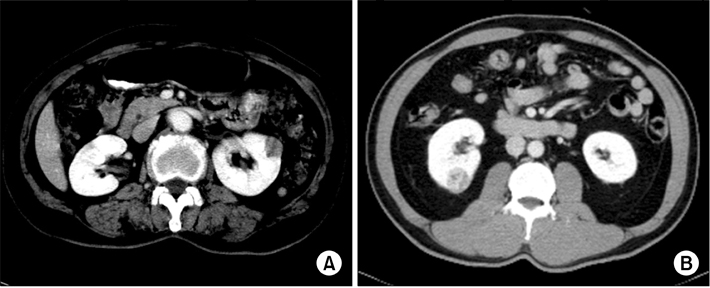
From January 2004 to March 2013, among 197 patients who underwent partial nephrectomy for suspicious renal cell carcinoma (RCC), the medical records of 153 patients with tumors (AML or RCC) < or =3 cm in diameter were retrospectively reviewed. Patient characteristics including age, gender, type of surgery, size and location of tumor, pathologic results, and specific findings of the imaging study ("ice-cream cone" shape) were compared between the AML and RCC groups.
RESULTS
AML was diagnosed in 18 patients and RCC was diagnosed in 135 patients. Gender (p=0.001), tumor size (p=0.032), and presence of the ice-cream cone shape (p=0.001) showed statistically significant differences between the AML group and the RCC group. In the multivariate logistic regression analysis, female gender (odds ratio [OR], 5.20; 95% confidence interval [CI], 1.45 to 18.57; p=0.011), tumor size (OR, 0.34; 95% CI, 0.12 to 0.92; p=0.034), and presence of the ice-cream cone shape (OR, 18.12; 95% CI, 4.97 to 66.06; p=0.001) were predictors of AML.
CONCLUSIONS
This study confirmed a high incidence of AML in females. Also, the ice-cream cone shape and small tumor size were significant predictors of AML in SRMs. These finding could be beneficial for counseling patients with SRMs.
MeSH Terms
Figure
Reference
-
1. Bosniak MA. The small (less than or equal to 3.0 cm) renal parenchymal tumor: detection, diagnosis, and controversies. Radiology. 1991; 179:307–317.2. Chow WH, Devesa SS. Contemporary epidemiology of renal cell cancer. Cancer J. 2008; 14:288–301.3. Pahernik S, Ziegler S, Roos F, Melchior SW, Thuroff JW. Small renal tumors: correlation of clinical and pathological features with tumor size. J Urol. 2007; 178:414–417.4. Remzi M, Ozsoy M, Klingler HC, Susani M, Waldert M, Seitz C, et al. Are small renal tumors harmless? Analysis of histopathological features according to tumors 4 cm or less in diameter. J Urol. 2006; 176:896–899.5. Schlomer B, Figenshau RS, Yan Y, Venkatesh R, Bhayani SB. Pathological features of renal neoplasms classified by size and symptomatology. J Urol. 2006; 176(4 Pt 1):1317–1320.6. Touijer K, Jacqmin D, Kavoussi LR, Montorsi F, Patard JJ, Rogers CG, et al. The expanding role of partial nephrectomy: a critical analysis of indications, results, and complications. Eur Urol. 2010; 57:214–222.7. Frank I, Blute ML, Cheville JC, Lohse CM, Weaver AL, Zincke H. Solid renal tumors: an analysis of pathological features related to tumor size. J Urol. 2003; 170(6 Pt 1):2217–2220.8. Lane BR, Babineau D, Kattan MW, Novick AC, Gill IS, Zhou M, et al. A preoperative prognostic nomogram for solid enhancing renal tumors 7 cm or less amenable to partial nephrectomy. J Urol. 2007; 178:429–434.9. Murphy AM, Buck AM, Benson MC, McKiernan JM. Increasing detection rate of benign renal tumors: evaluation of factors predicting for benign tumor histologic features during past two decades. Urology. 2009; 73:1293–1297.10. Glassman D, Chawla SN, Waldman I, Johannes J, Byrne DS, Trabulsi EJ, et al. Correlation of pathology with tumor size of renal masses. Can J Urol. 2007; 14:3616–3620.11. Thompson RH, Kurta JM, Kaag M, Tickoo SK, Kundu S, Katz D, et al. Tumor size is associated with malignant potential in renal cell carcinoma cases. J Urol. 2009; 181:2033–2036.12. Marhuenda A, Martin MI, Deltoro C, Santos J, Rubio Briones J. Radiologic evaluation of small pretreatment management. Adv Urol. 2008; 415848.13. Verma SK, Mitchell DG, Yang R, Roth CG, O'Kane P, Verma M, et al. Exophytic renal masses: angular interface with renal parenchyma for distinguishing benign from malignant lesions at MR imaging. Radiology. 2010; 255:501–507.14. Wagner BJ, Wong-You-Cheong JJ, Davis CJ Jr. Adult renal hamartomas. Radiographics. 1997; 17:155–169.15. Jinzaki M, Tanimoto A, Narimatsu Y, Ohkuma K, Kurata T, Shinmoto H, et al. Angiomyolipoma: imaging findings in lesions with minimal fat. Radiology. 1997; 205:497–502.16. Kim JK, Park SY, Shon JH, Cho KS. Angiomyolipoma with minimal fat: differentiation from renal cell carcinoma at biphasic helical CT. Radiology. 2004; 230:677–684.17. Bosniak MA, Megibow AJ, Hulnick DH, Horii S, Raghavendra BN. CT diagnosis of renal angiomyolipoma: the importance of detecting small amounts of fat. AJR Am J Roentgenol. 1988; 151:497–501.18. Israel GM, Hindman N, Hecht E, Krinsky G. The use of opposed-phase chemical shift MRI in the diagnosis of renal angiomyolipomas. AJR Am J Roentgenol. 2005; 184:1868–1872.19. Outwater EK, Bhatia M, Siegelman ES, Burke MA, Mitchell DG. Lipid in renal clear cell carcinoma: detection on opposed-phase gradient-echo MR images. Radiology. 1997; 205:103–107.20. Duchene DA, Lotan Y, Cadeddu JA, Sagalowsky AI, Koeneman KS. Histopathology of surgically managed renal tumors: analysis of a contemporary series. Urology. 2003; 62:827–830.21. Filipas D, Fichtner J, Spix C, Black P, Carus W, Hohenfellner R, et al. Nephron-sparing surgery of renal cell carcinoma with a normal opposite kidney: long-term outcome in 180 patients. Urology. 2000; 56:387–392.22. McKiernan J, Yossepowitch O, Kattan MW, Simmons R, Motzer RJ, Reuter VE, et al. Partial nephrectomy for renal cortical tumors: pathologic findings and impact on outcome. Urology. 2002; 60:1003–1009.23. Shannon BA, Cohen RJ, de Bruto H, Davies RJ. The value of preoperative needle core biopsy for diagnosing benign lesions among small, incidentally detected renal masses. J Urol. 2008; 180:1257–1261.24. Volpe A, Mattar K, Finelli A, Kachura JR, Evans AJ, Geddie WR, et al. Contemporary results of percutaneous biopsy of 100 small renal masses: a single center experience. J Urol. 2008; 180:2333–2337.25. Volpe A, Kachura JR, Geddie WR, Evans AJ, Gharajeh A, Saravanan A, et al. Techniques, safety and accuracy of sampling of renal tumors by fine needle aspiration and core biopsy. J Urol. 2007; 178:379–386.26. Schmidbauer J, Remzi M, Memarsadeghi M, Haitel A, Klingler HC, Katzenbeisser D, et al. Diagnostic accuracy of computed tomography-guided percutaneous biopsy of renal masses. Eur Urol. 2008; 53:1003–1011.27. Rendon RA. Active surveillance as the preferred management option for small renal masses. Can Urol Assoc J. 2010; 4:136–138.


